Open Craniosynostosis Surgery

Treatments for Craniosynostosis

At El Paso Craniofacial Team, the primary treatment for craniosynostosis is surgery. First performed in the late 1800s, surgical approaches have continued to evolve and improve. The two main approaches are Calvarial Vault Remodel (CVR) and endoscopic-assisted surgery, with each tailored to the child’s age, the affected suture, and the severity of the condition.

Calvarial Vault Remodel (CVR)

CVR is the traditional method, most often used in children older than 10–12 months. It involves:

Wide exposure of the skull

Removal of large sections of bone

Reshaping of the deformed bone

Replacing and fixation of the bone to achieve a more typical skull shape

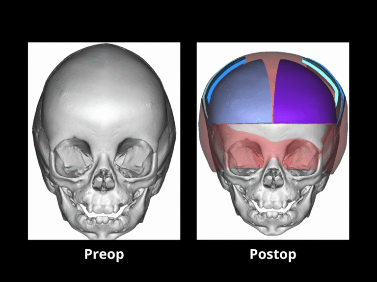
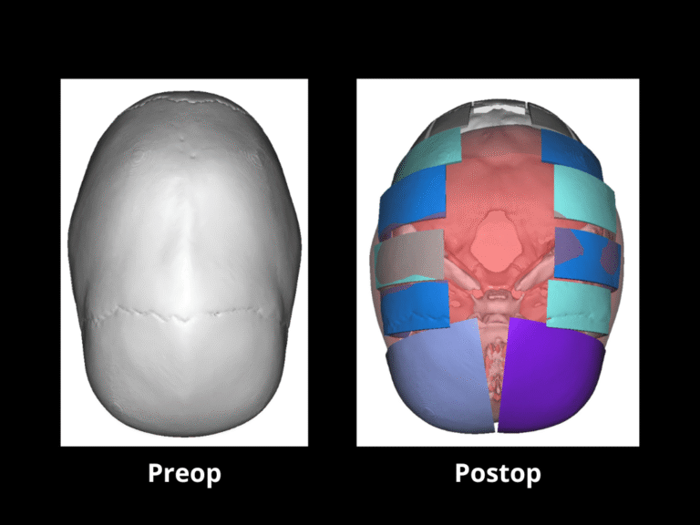
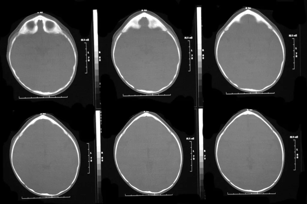
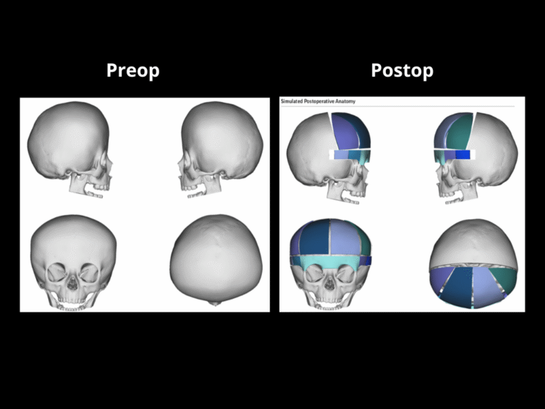
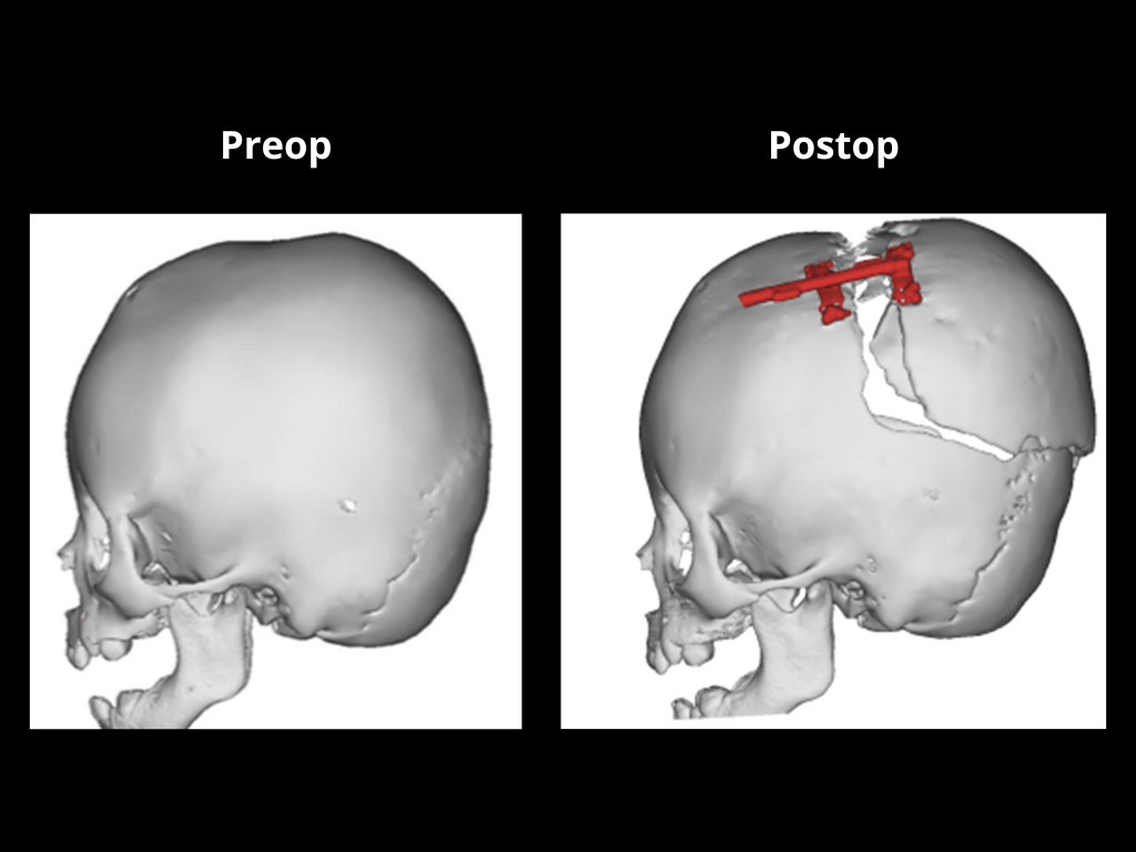
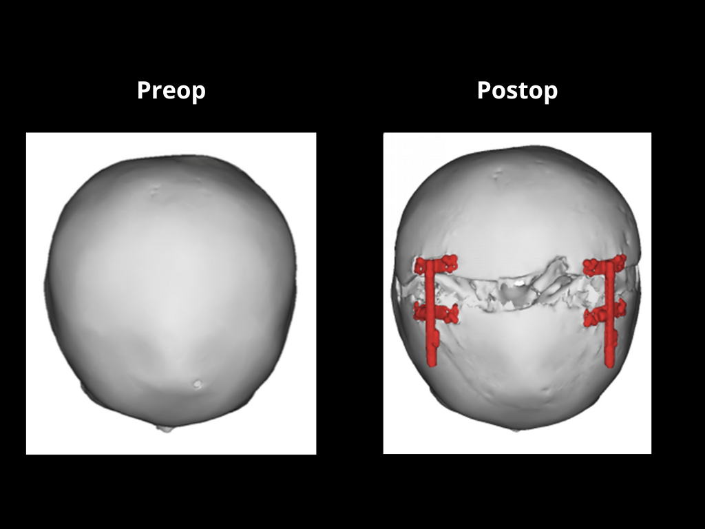
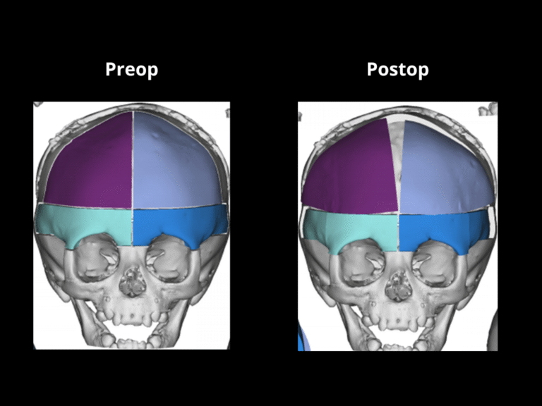
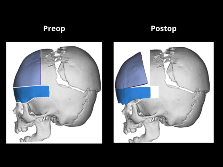
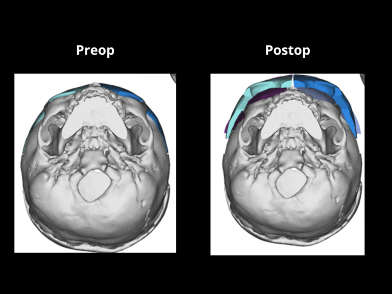
While CVR provides immediate results, the procedure is lengthy (3–9 hours), often requires transfusions, and involves longer recovery times.

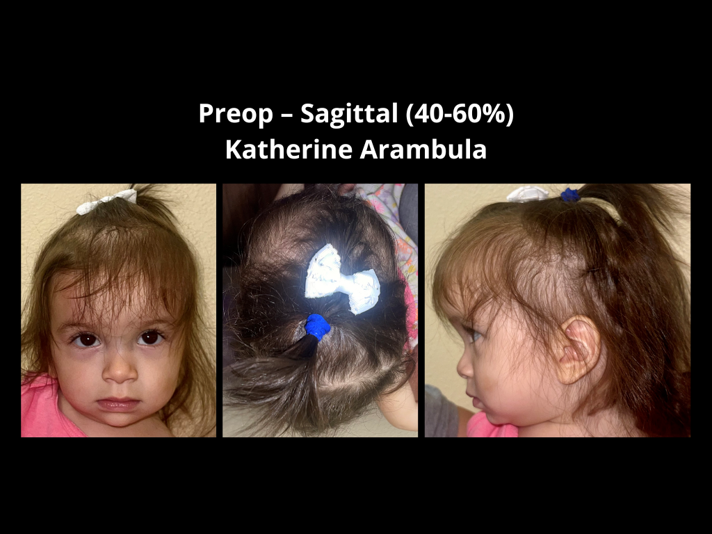
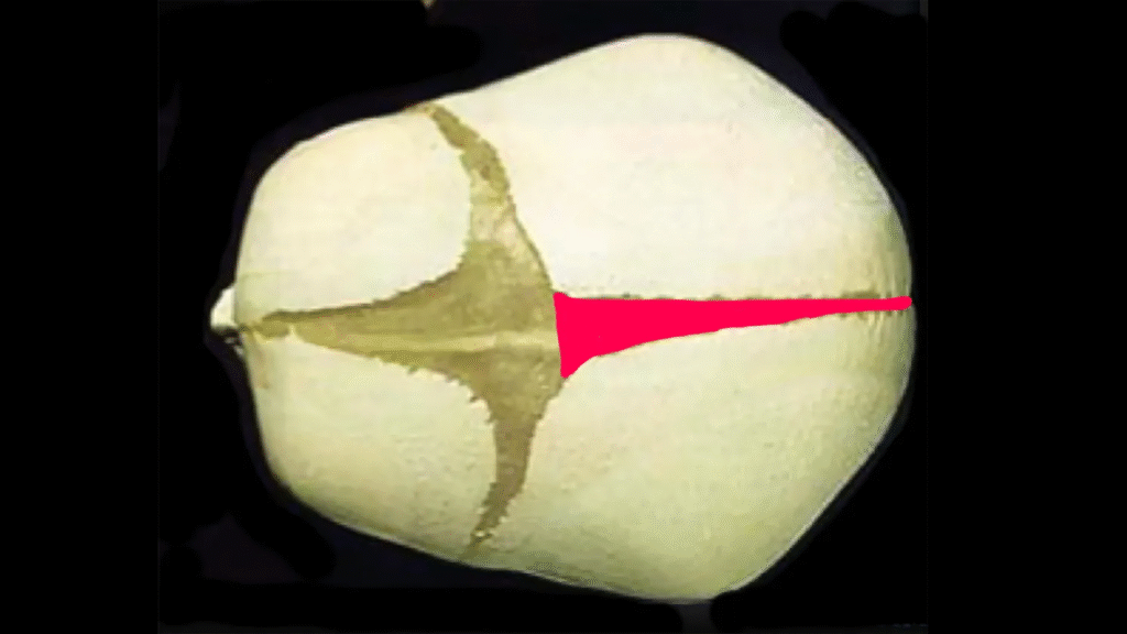
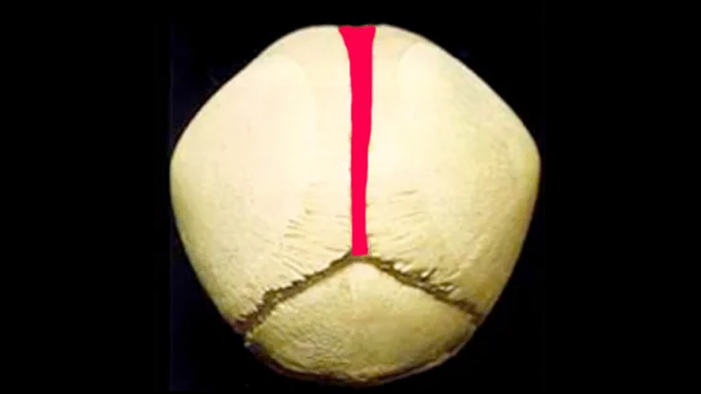
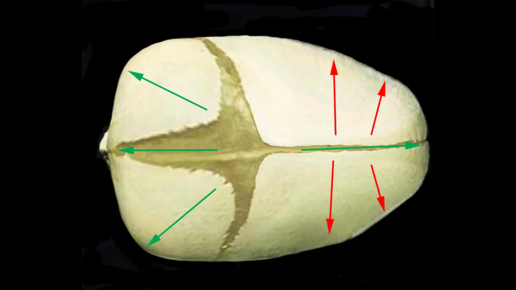
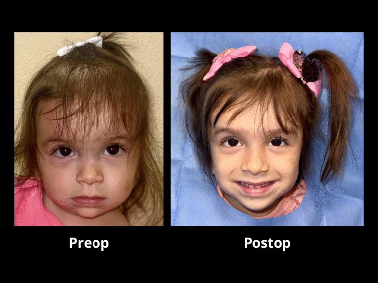
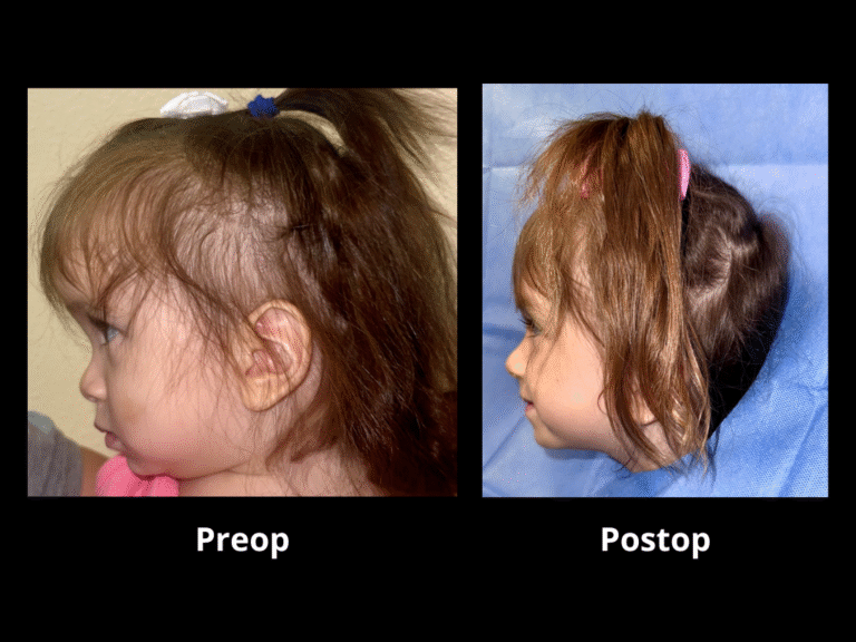
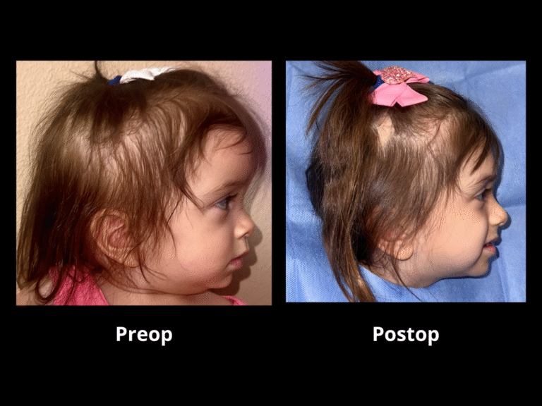
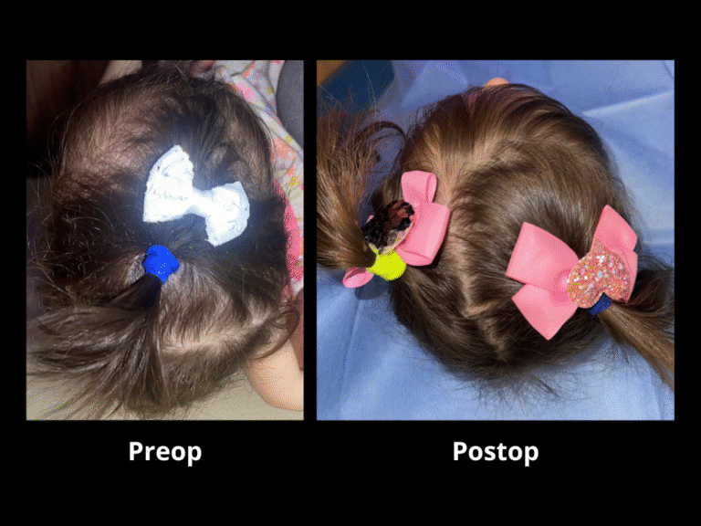
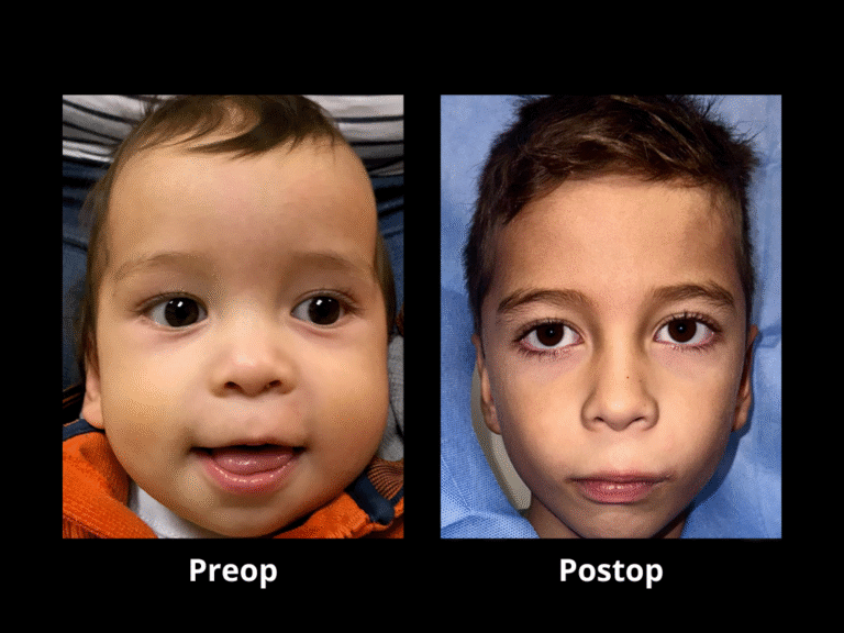
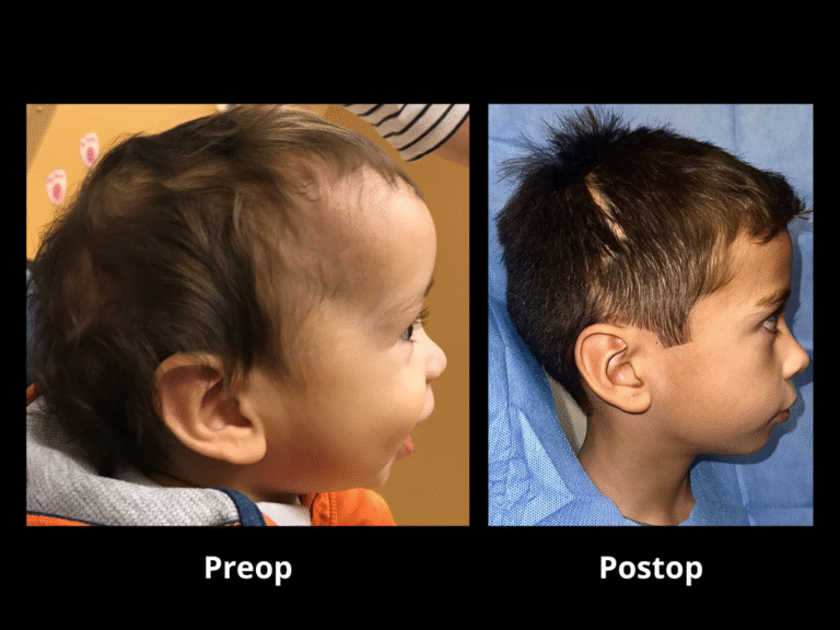
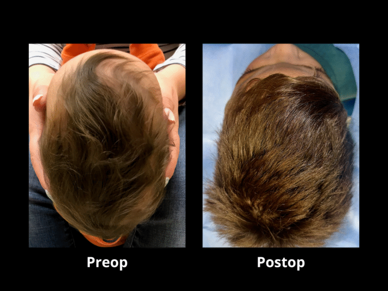
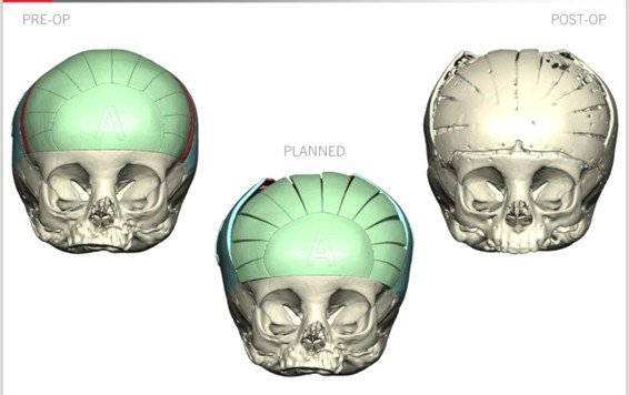
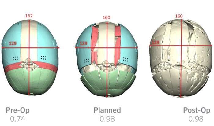
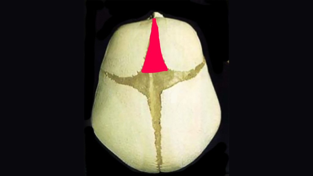
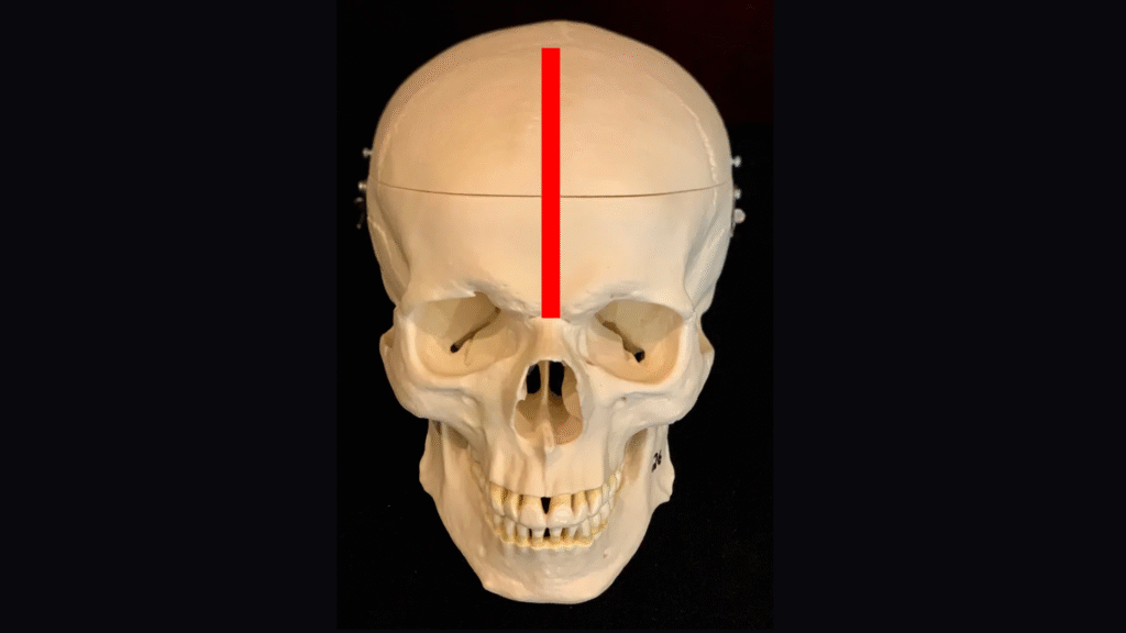
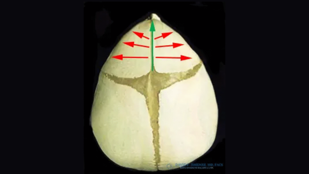
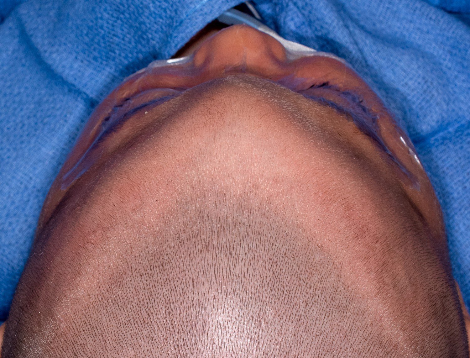
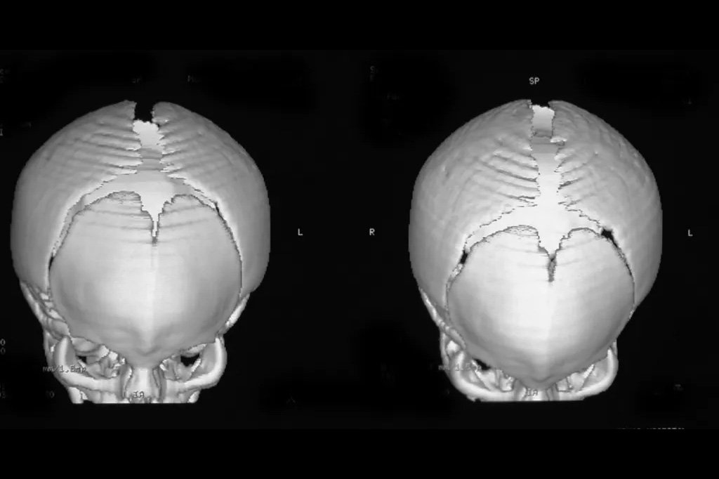
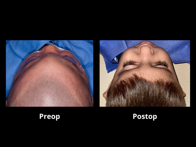
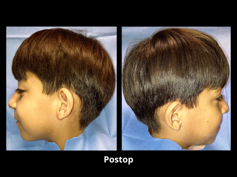
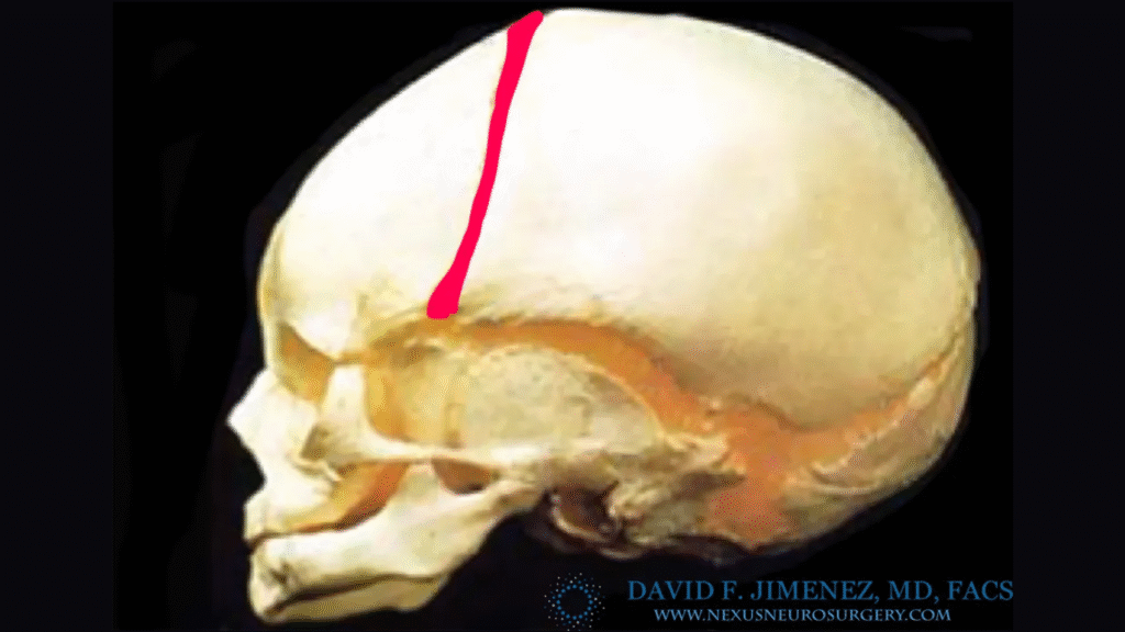
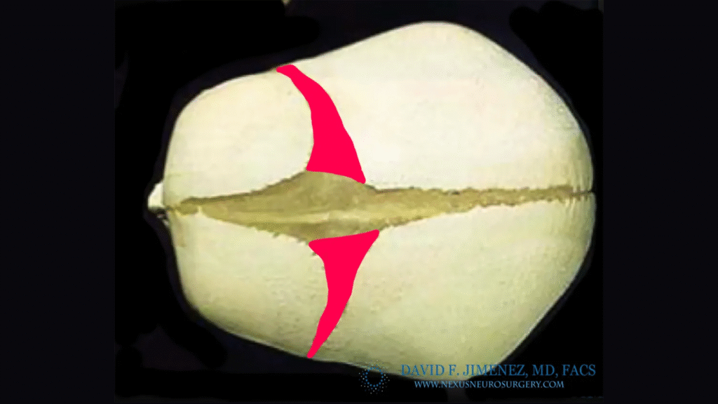
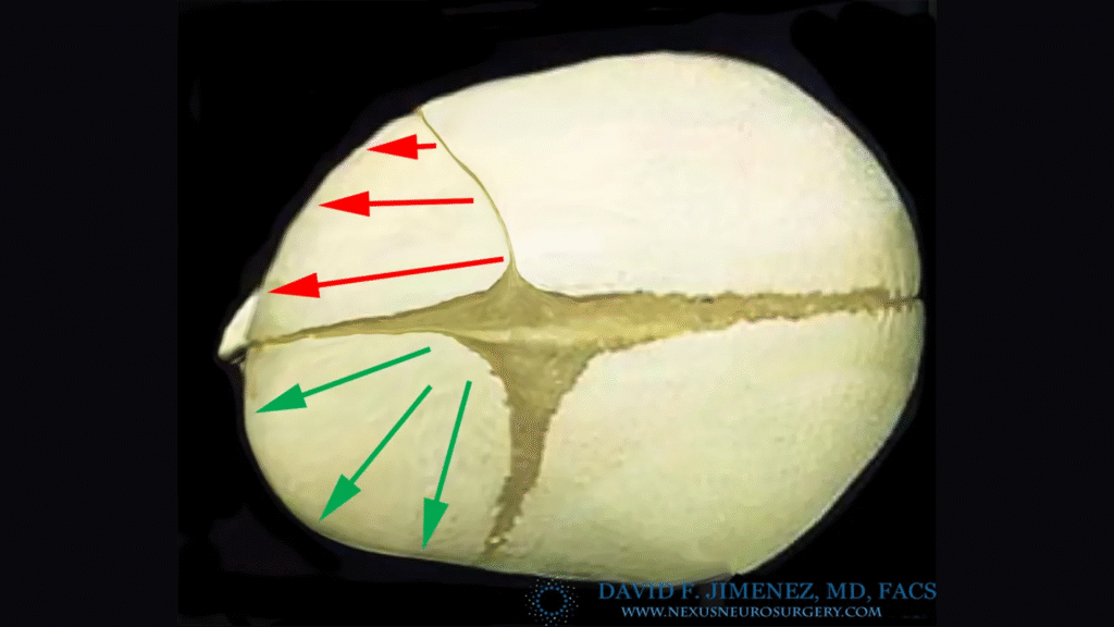
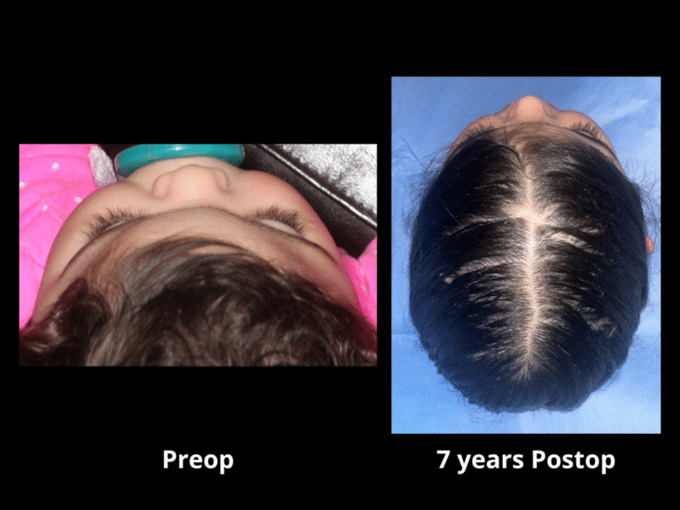
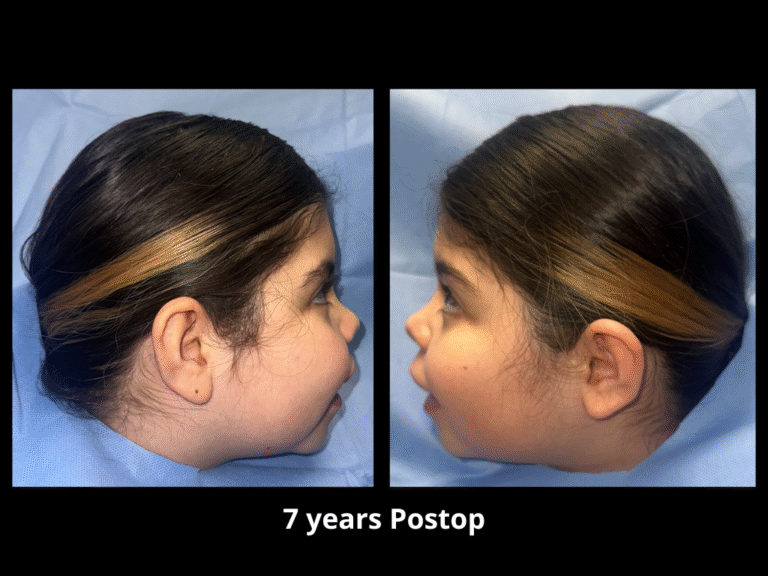
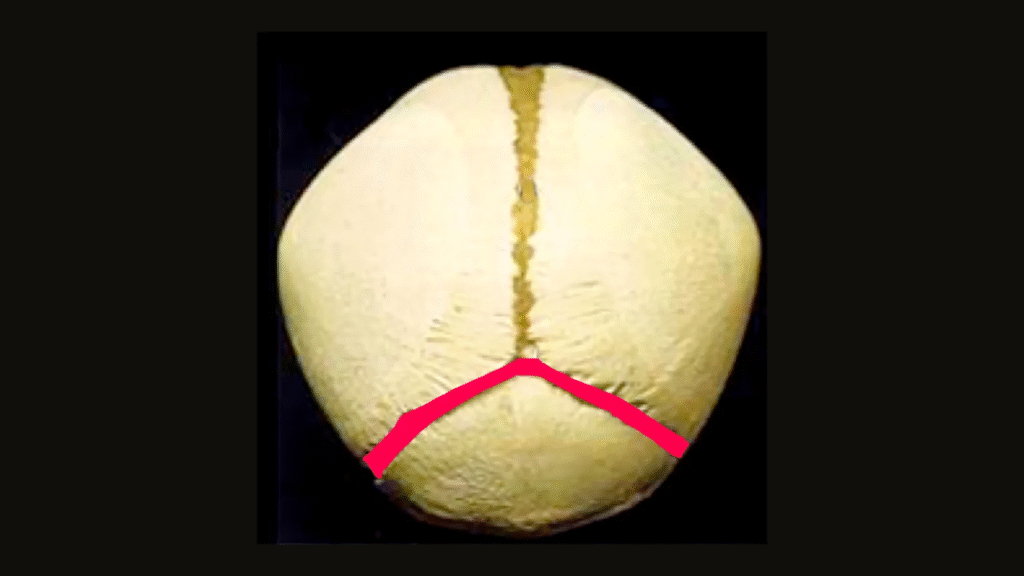
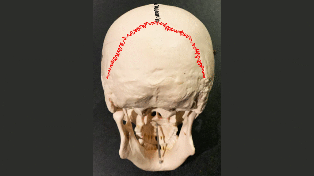
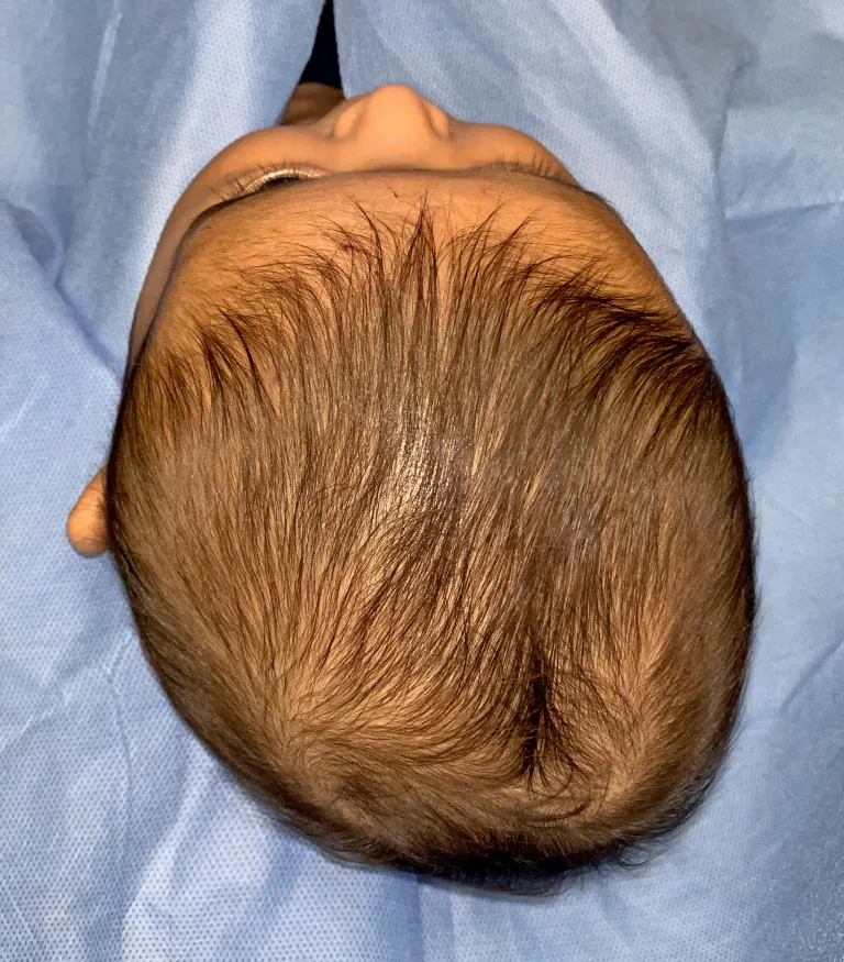
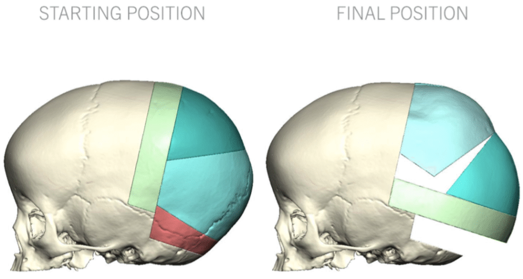
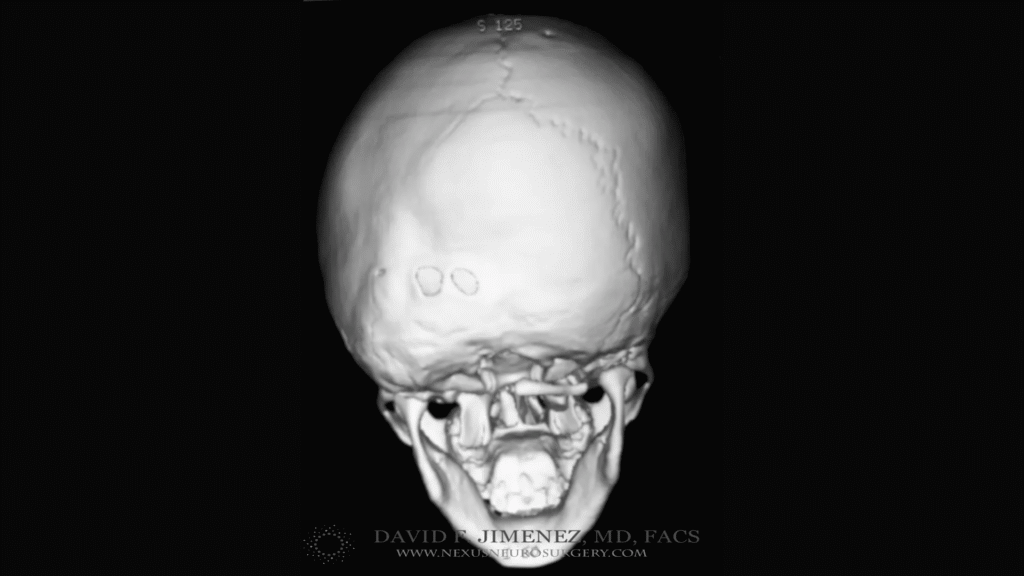
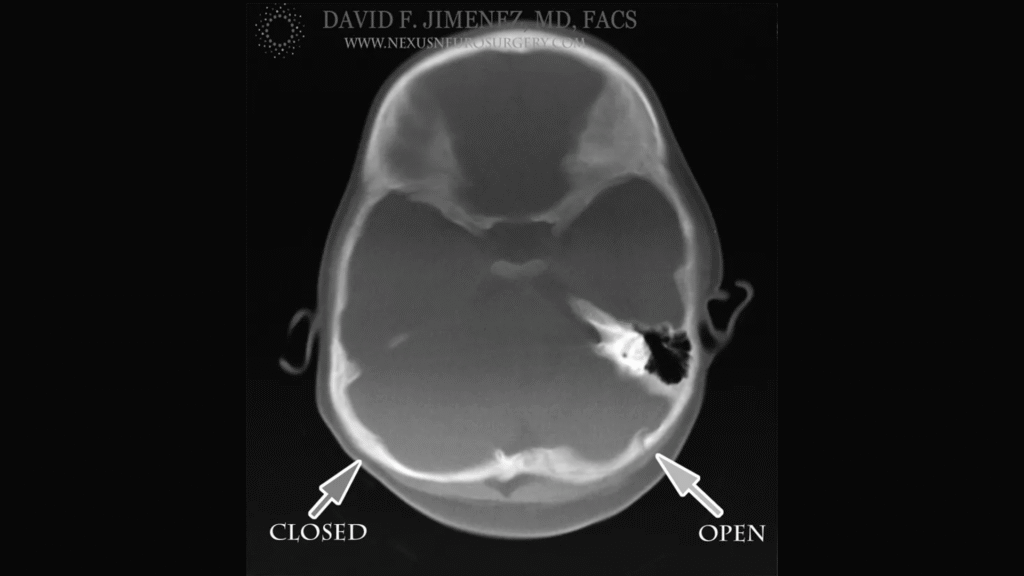
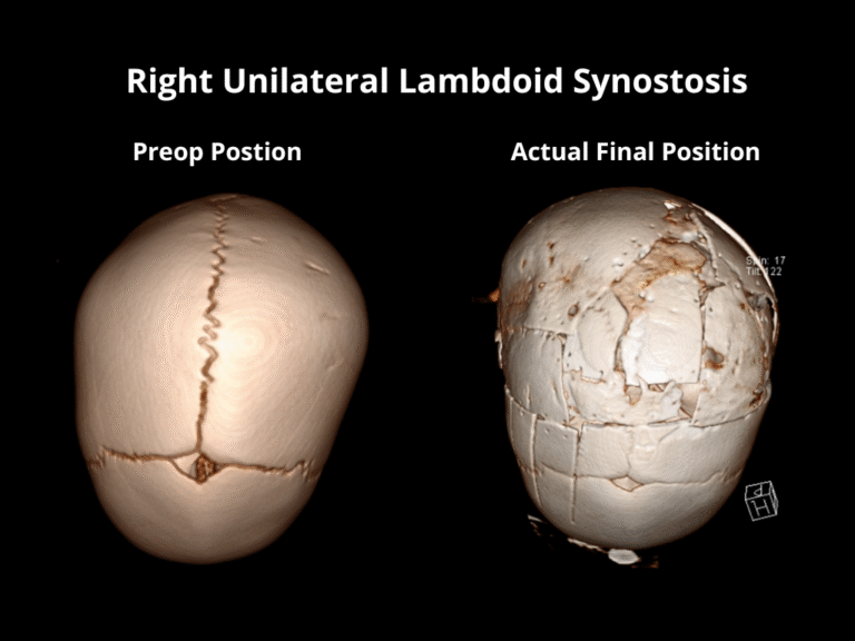
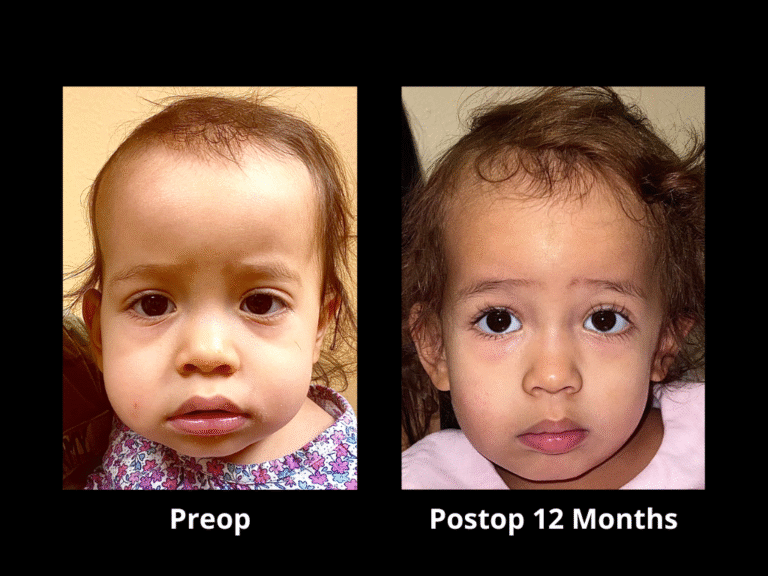
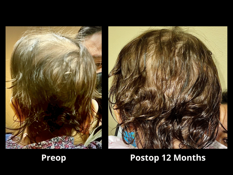
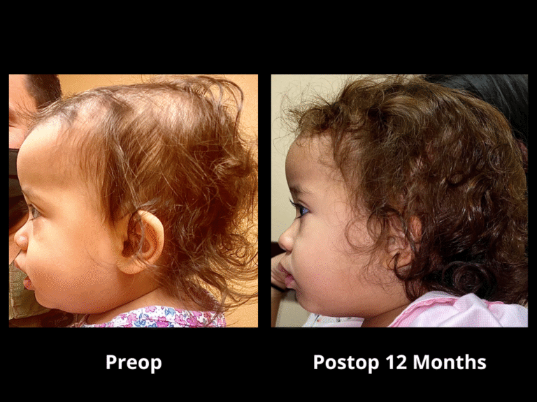
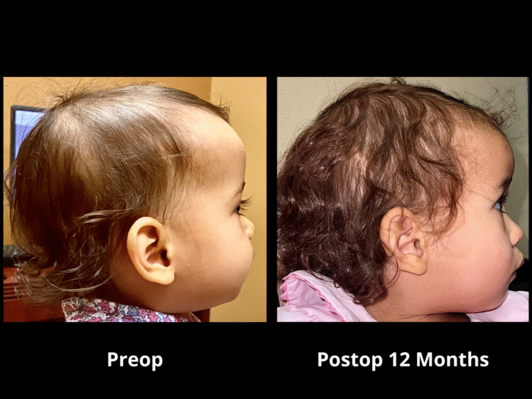
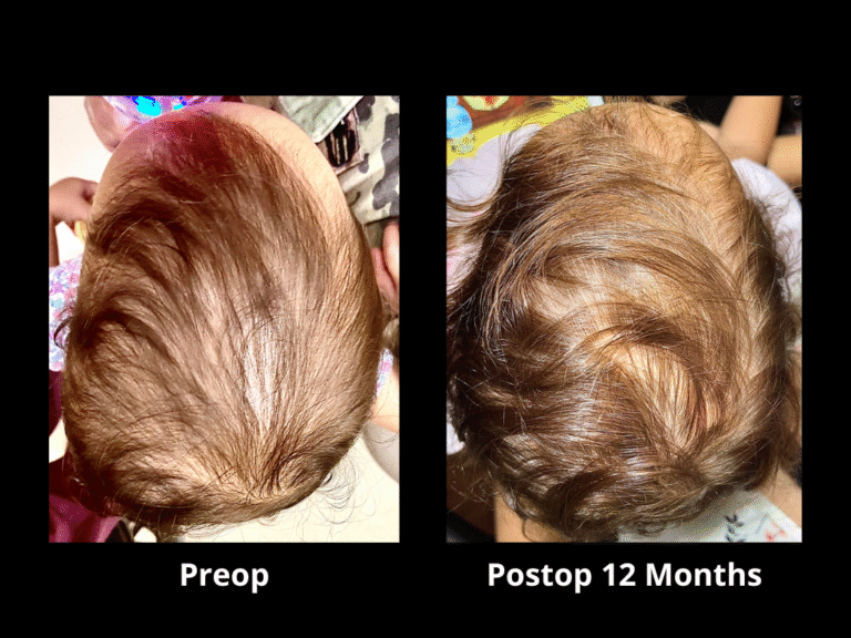
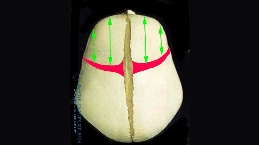
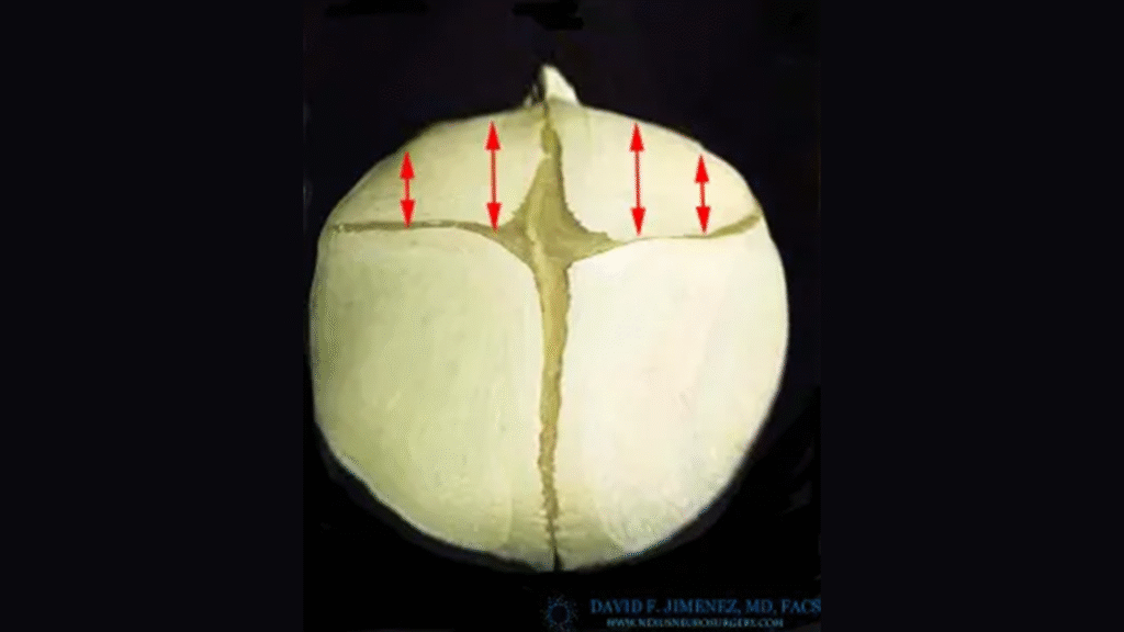
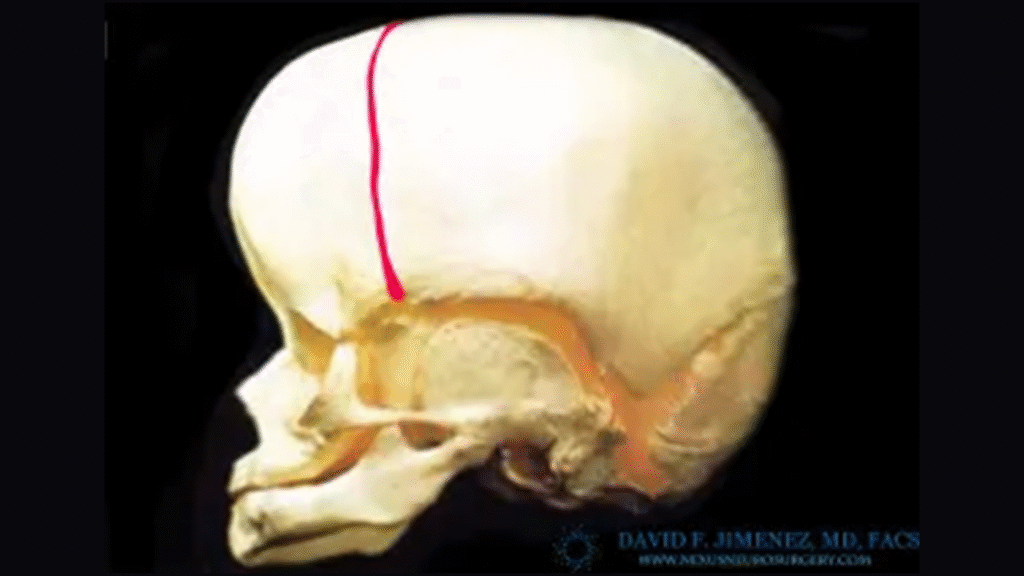
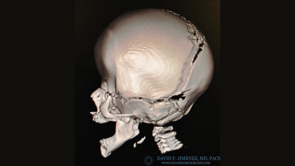
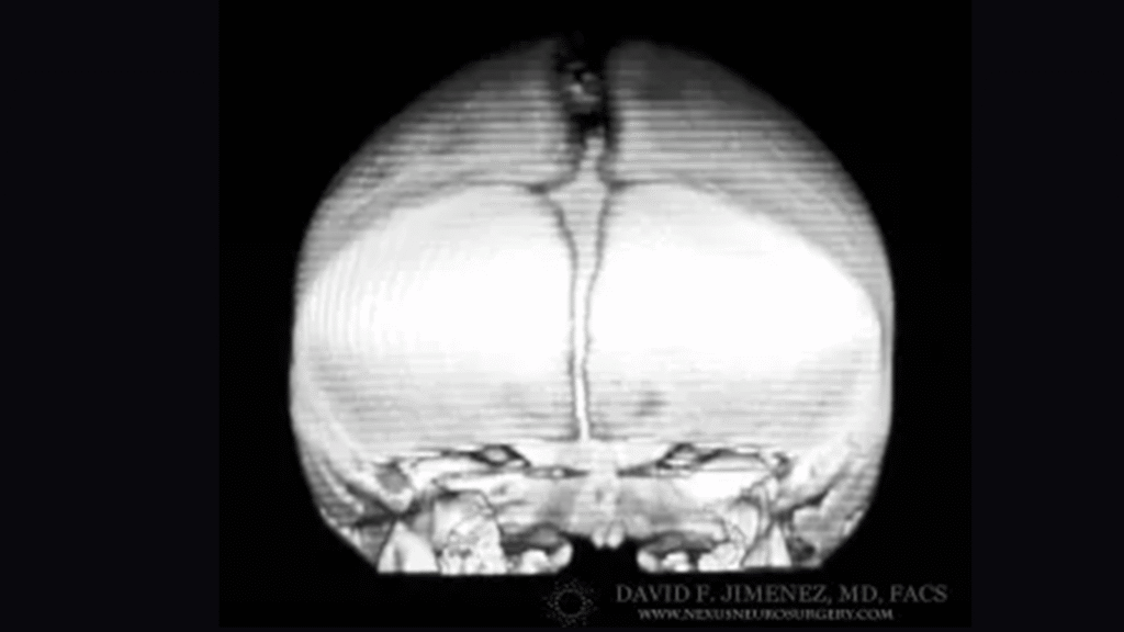
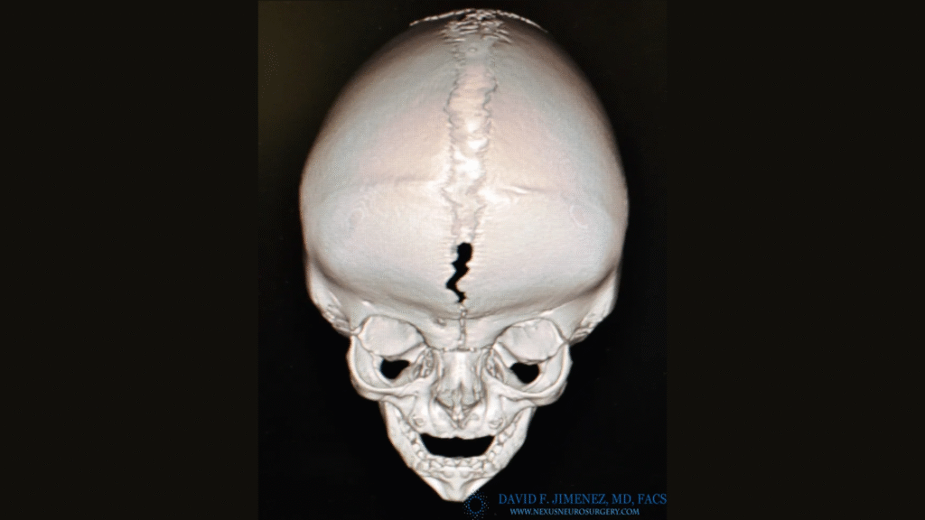
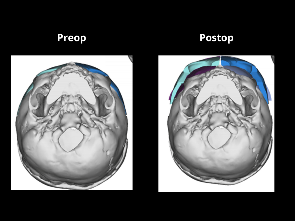
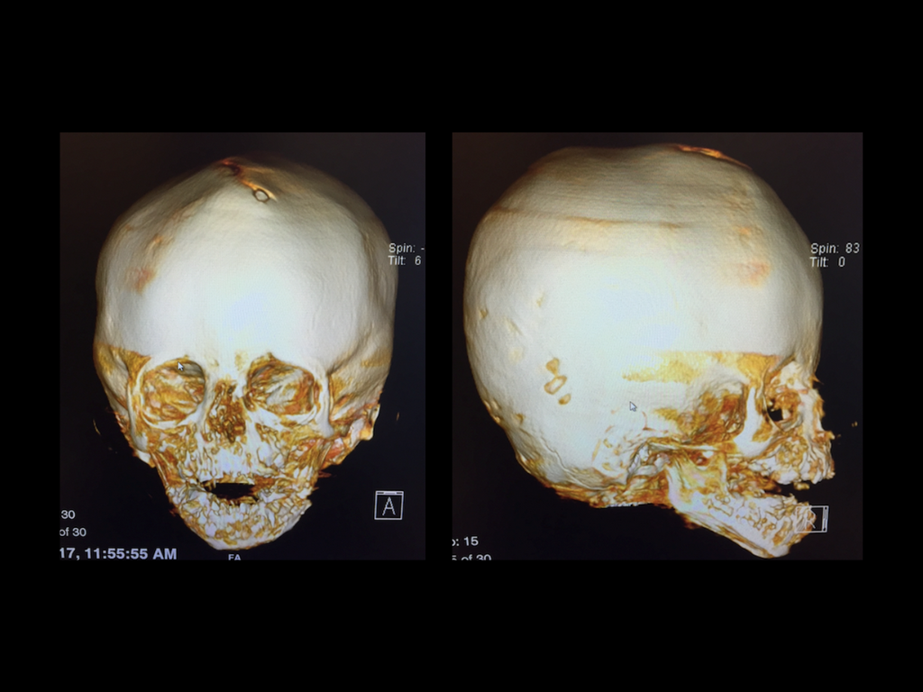
Recognizing these challenges, El Paso Craniofacial Team pioneered the use of endoscopic-assisted surgery more than two decades ago. This minimally invasive technique is performed in very young infants, ideally before 12 weeks of age. Through small incisions, the fused suture is released, allowing the brain’s rapid growth to remodel the skull naturally. Postoperative helmet therapy then helps guide growth into a normal shape.

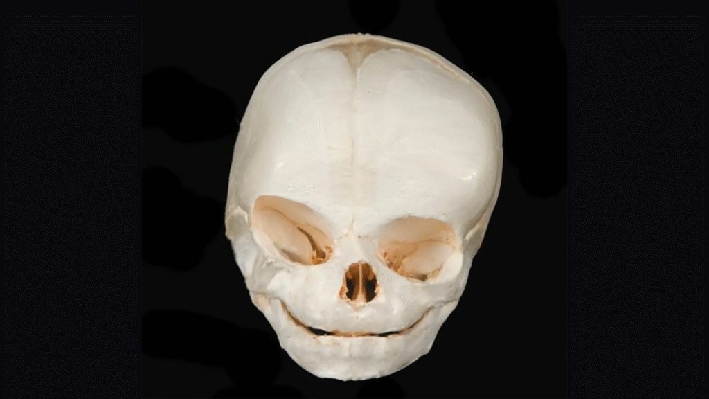
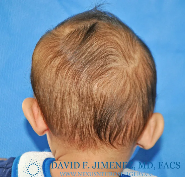
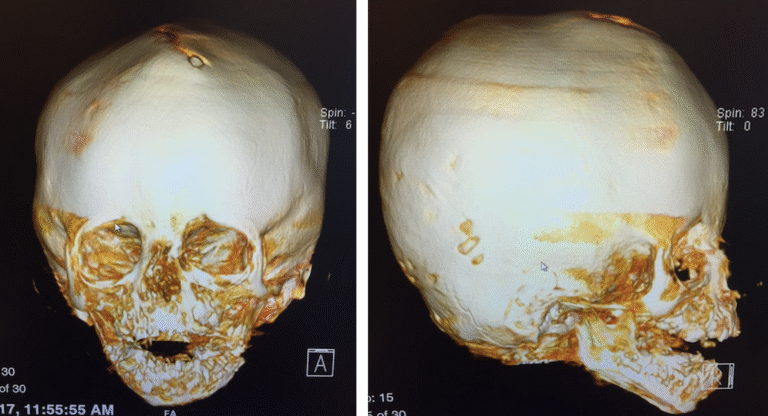
Custom helmets, manufactured by FDA-cleared Orthomerica Products, Inc., are adjusted regularly to accommodate rapid growth. Helmets do not restrict brain development; instead, they redirect growth safely and effectively.

Sagittal Synostosis

The sagittal suture runs along the midline of the skull, from the soft spot to the back of the head. When it fuses too early, the skull grows long and narrow in a shape called scaphocephaly.

The sagittal suture is located in the midline and behind the soft spot as demonstrated by the red area.

Back view of a normal newborn skull shows the back extension of the sagittal suture as it reaches the lambdoid sutures.

Upon premature closure of the suture, the skull cannot grow in the direction of the red arrows. The normal brain grows in the path of the other open sutures and of least resistance as shown by the green arrows.

Sagittal Synostosis

The classical presentation consists of a narrow and elongated skull with very prominent and protruding forehead. Additionally, the back of the head becomes pointed and tapered. This condition is known as scaphocephaly. However, the head may look different depending on whether the entire suture is closed or only parts of it and when the closure occurs. Additionally, the head can become tall and narrow.

Sagittal Synostosis

Before & After

Sagittal Synostosis

Appropriate Planning and Reconstruction

Demonstrating a preoperative skull with detailed surgical markings for planned craniectomies.
The sequence illustrates the transition from planned expansion to the postoperative expansion achieved, showcasing precise surgical planning and successful cranial reconstruction outcomes.

Metopic Synostosis

The metopic suture extends from the soft spot down to the root of the nose. Premature closure leads to a triangular forehead shape known as trigonocephaly, often accompanied by closely spaced eyes (hypotelorism) and angled eyebrows.

The metopic suture is located in the midline and in front the soft spot as demonstrated by the red area.

The metopic suture extends from the soft spot all the way down to the root of the nose (nasofrontal suture) in the area between the eyes.

The premature closure of the metopic suture prevents the front center of the skull from moving sideways and the front sides from  moving forwards (red arrows) The midline moves forward ( green arrow) causing a midline ridge and the classical triangular shaped head.

Metopic Synostosis

The classical presentation consists of a narrow and elongated skull with very prominent and protruding forehead. Additionally, the back of the head becomes pointed and tapered. This condition is known as scaphocephaly. However, the head may look different depending on whether the entire suture is closed or only parts of it and when the closure occurs. Additionally, the head can become tall and narrow.

Metopic Synostosis

Radiology

Metopic synostosis is a clinical diagnosis, meaning that it is made by examining the patient and identifying the associated deformation of the head and face. CT  scans and X rays are not necessary to make the diagnosis. However, the images clearly show the skull changes related to this condition.

Metopic Synostosis

Post Operative Swelling

After surgery, patients commonly experience facial swelling and mild bruising, especially around the eyes. This is a normal part of the healing process and tends to peak within the first 24 hours. The eyes may appear puffy or partially swollen shut, but this gradually improves over the next 48–72 hours. Discomfort during the first few hours is usually well controlled with Tylenol and Motrin. By the next morning, most patients are smiling, feeding well, and returning to their usual routines. 

Metopic Synostosis

Before & After

Coronal Synostosis

The coronal  sutures are located on either side of  the head and adjoin the soft spot in the middle. Their function is to allow the frontal lobes of the brain to grow and move forward. This in turn allows the forehead, eyes, eyebrows and nose to also move forward and downward. Premature closure results in a number of deformities related to these structures’ inability to advance normally.

Side view of the newborn skull shows the location of the left coronal suture highlighted in red.

Top view of the skull depicts the  location of the coronal sutures on either side of the soft spot and extending to the sides.

The premature closure of the right coronal suture prevents the front right side of the skull from moving forwards (red arrows) leading to flattening of the right side of the forehead. The brain takes the path of least resistance and begins to grow  more on the left side (green arrows) leading to frontal bossing of the right.

Front view of a newborn skull with right coronal synostosis. The right eye socket is elevated and pulled upwards giving the appearance on skull x-rays of the so called “harlequin eye”.

The nose is pushed to the opposite left side (green arrow), the right eye socket is elevated and moves higher than the left one (red arrow), the right side of the face is pushed inwards (blue arrows) while the left  side of the forehead is pushed outwards (yellow arrows).

Coronal Synostosis

The classical presentation consists of coronal craniosynostosis consists of:

1. Elevation of the eye on the affected side (vertical dystopia)

2. Deviation of the nose to the opposite side

3. Flattening of the forehead on the affected side (frontal plagiocephaly)

4. Protruding eye on the affected side (proptosis)

5.  Overgrowth of forehead on the opposite side.

6.  Head deviates to the side (cranial scoliosis)

Two month old male with significant cranial-facial deviation (scoliosis) as evidenced by the angulation of a line going from the root of the nose to the mid chin and top of the head as seen in coronal synostosis

Cranial scoliosis (red line) and vertical dystopia (left eye elevated above right eye) as demonstrated by the black line in infant girl with left coronal synostosis.

Coronal Synostosis

Before & After

Lambdoid Synostosis

The lambdoid suture runs along the back of the skull, extending from the midline to behind the ears. Lambdoid synostosis is the rarest form of single-suture craniosynostosis.

The lambdoid suture is located in the back of the skull as seen on this newborn’s skull and marked with red.

Both lambdoid sutures, marked in red, as seen on the back of a newborn’s skull.

Back view of an adult skull showing the location of both lambdoid sutures (red lines) as the extend from the mastoid bones to the sagittal suture (black lines) on the top of the skull.

Lambdoid Synostosis

The classical presentation consists of flattening and depression at the back of the head on the affected side. From a top view, the skull may appear trapezoidal in shape. From behind, the mastoid region and ear on the affected side are often displaced downward compared to the unaffected side. The head also tends to tilt laterally, producing a form of cranial scoliosis. It is important to distinguish this condition from head shape changes caused by positional molding, which commonly occur in infants who sleep on their backs as recommended by safe sleep guidelines.

Lambdoid Synostosis

Right Unilateral Lambdoid Synostosis

Lambdoid Synostosis

Radiology

Unlike other single suture synostosis, radiologic studies are needed to assure that the correct diagnosis is made. Because it can be easily confused with positional molding, X-rays  or CT scans are very useful in helping make the diagnosis.

3-D CT scan of patient with partial closure of the left lambdoid suture near the midline sagittal suture.(Black arrows)

3-D CT scan of infant with an area of “spot welding” in the middle of the right lambdoid suture. Notice that even this very small amount of  synostosis causes significant deformation of the skull base as seen with downward displacement of the right mastoid bone (B) in relation to the normal mastoid bone (A).

3-D CT scan of patient with premature closure of most of the left lambdoid suture.

CT scan showing thin slices of the skull and a closed left lambdoid suture.

Arrow on the left shows the closed suture whereas the arrow on the right shows an open suture on the other side.

3-D CT scan shows the downward displacement of the mastoid bone in this child with left lambdoid synostosis.

Lambdoid Synostosis

Right Unilateral Lambdoid Synostosis

Lambdoid Synostosis

Before & After

Bicoronal Non-Syndromic Synostosis

Non-syndromic bicoronal synostosis occurs when both coronal sutures close prematurely without being linked to genetic syndromes such as Apert or Crouzon. These children are otherwise normal, with no developmental delays or systemic conditions.

Open coronal sutures (red areas) allow both frontal lobes to grow and advance forward (green arrows).

Closed coronal sutures keep the frontal  lobes from moving forward and cause a shallow and foreshortened frontal cranial vault (red arrows).

Normal brain growth allows the bones around the eyes and face to move forward.

Premature bicoronal synostosis keeps the bones from moving forward and causes the mid face and eye sockets to be recessed and the forehead to bulge abnormally.

Bicoronal Non-Syndromic Synostosis

The patient’s head, is wide and shallow (brachycephaly). The eyes protrude in front of the eye brows and eye sockets (proptosis). The side of the eyes tend to angulate downward and because the brain can’t grow forward, it grows tall causing the forehead and cranial vault to be high ( turricephaly). The patients are otherwise normal with no evidence of fused fingers or toes (syndactilty), wide and broad thumbs or fused elbows.

Radiology

CT scanning is the gold standard test for making the appropriate diagnosis. The closed sutures are very evident as are all of the other aforementioned clinical findings. MRI scans are used as an adjunct to better visualize the brain and intracranial structures, if indicated.

Bicoronal Non-Syndromic Synostosis

Before & After

Multi-suture Craniosynostosis

Multi-suture craniosynostosis is a condition in which two or more cranial sutures fuse prematurely. This restricts normal skull growth perpendicular to the affected sutures, forcing the skull to grow abnormally in other directions. It often leads to severe cranial deformities and, in some cases, increased intracranial pressure (ICP).

Multi-suture Craniosynostosis

EPCH Cranial and Facial Surgery Team Santos.
He wants to become a Craniofacial Surgeon.

Pansynostosis

Pansynostosis (All Sutures Fused)
Open cranial approach addressing bilambdoid, bicoronal, sagittal, and metopic suture fusion to restore cranial volume and symmetry.

Posterior vault distraction (15 mm) followed by anterior cranial vault reconstruction to restore skull volume and contour.

After 25 days of Distraction – Distractor Arms in Place.

Copper-Beaten Skull Appearance Secondary to increases in Intracranial Pressure

Pansynostosis occurs when all cranial sutures close prematurely, restricting skull growth in every direction. This condition can lead to increased intracranial pressure, abnormal head shape, and potential developmental delays. Early surgical correction helps restore skull volume and allows for normal brain growth.

Begin Your Child’s Treatment Journey

Craniosynostosis, whether sagittal, metopic, bicoronal, or lambdoid, can be successfully treated when identified early. El Paso Craniofacial Team offers advanced, minimally invasive surgical care combined with cranial helmet therapy to ensure safe, effective, and lasting outcomes for children and their families.