Endoscopic Treatment for Craniosynostosis

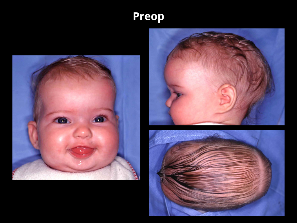
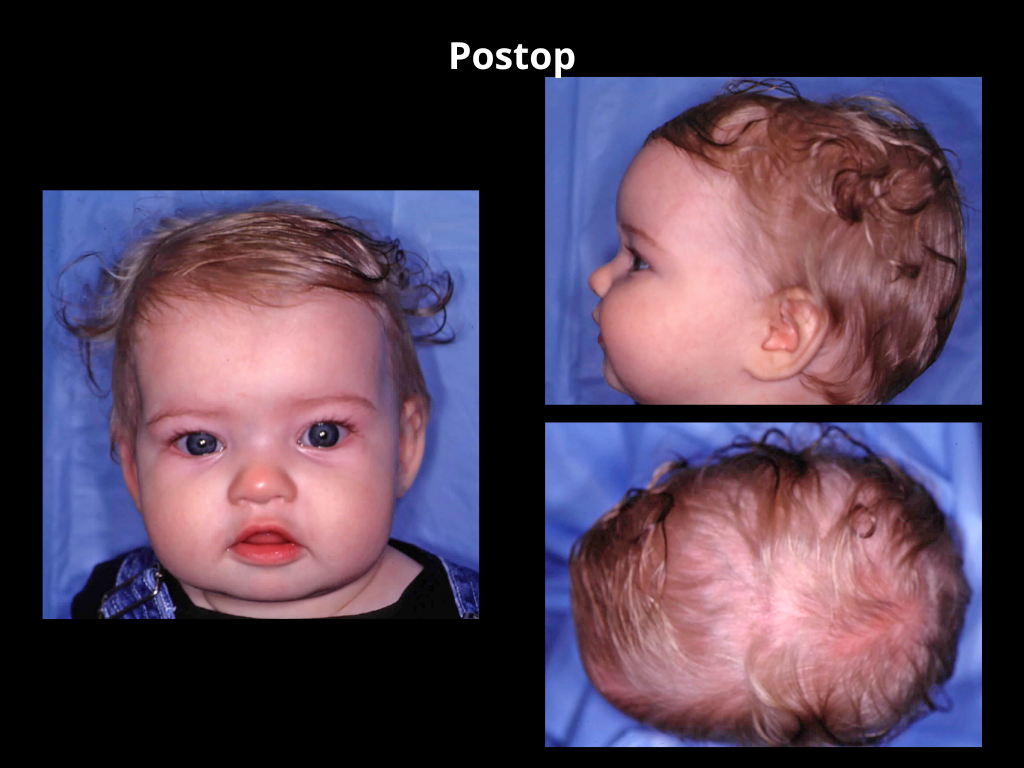
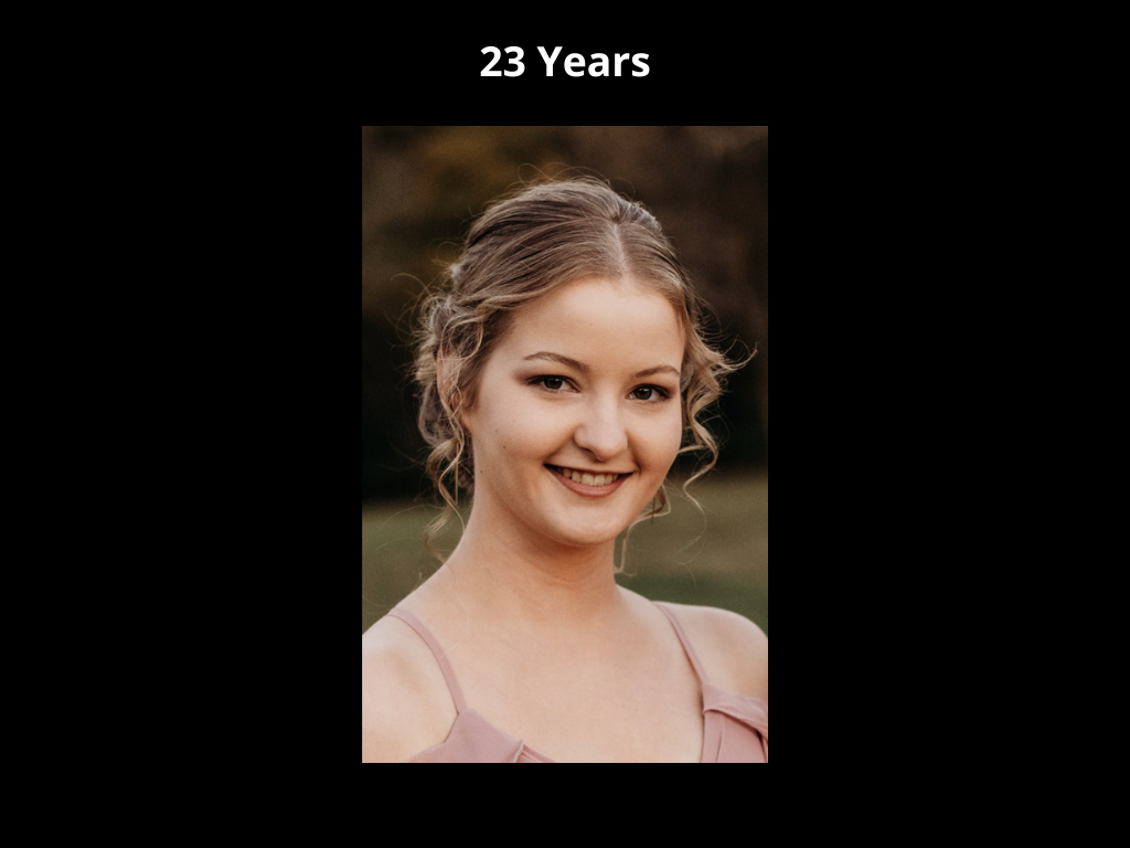
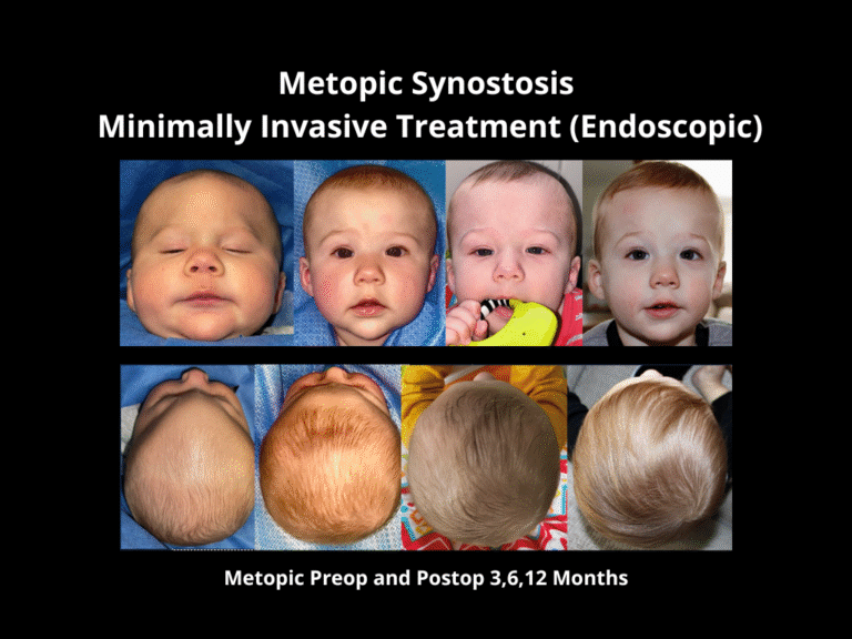
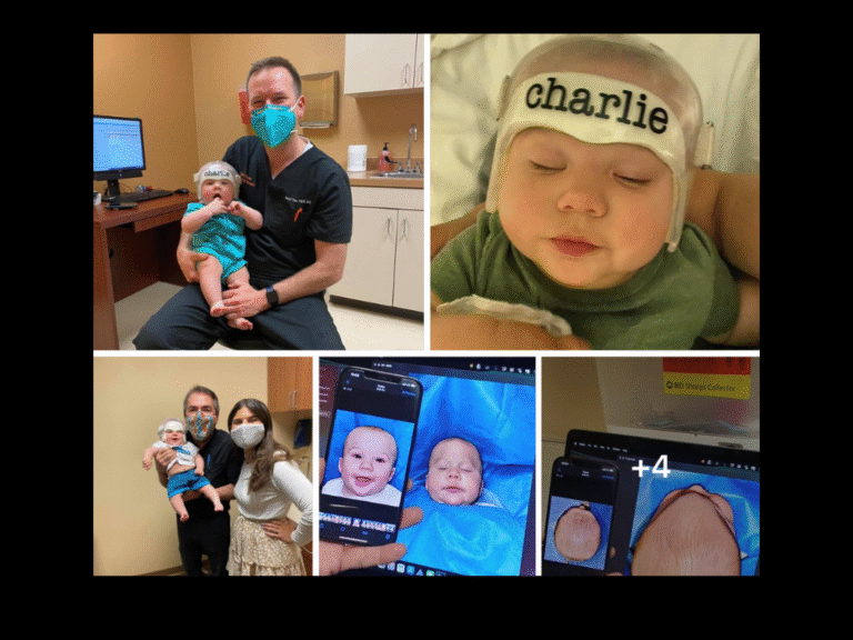
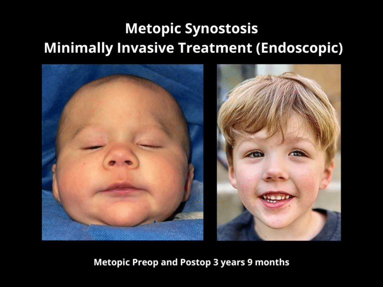
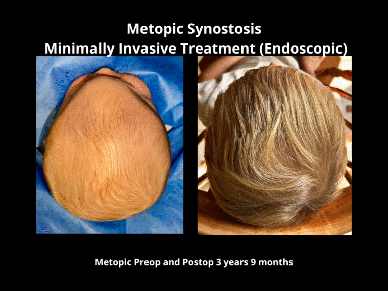
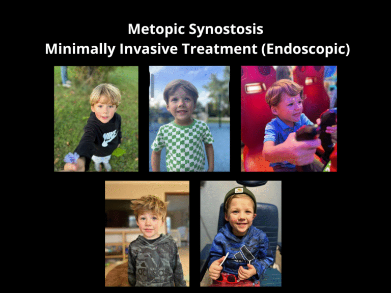
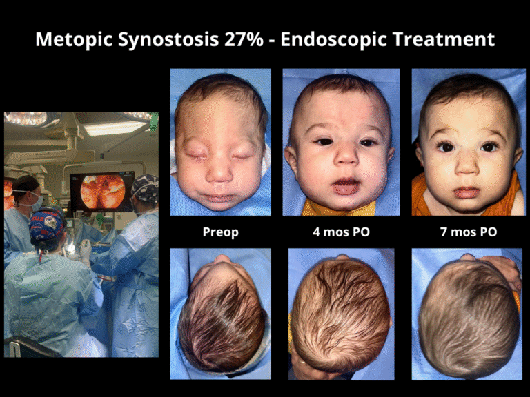
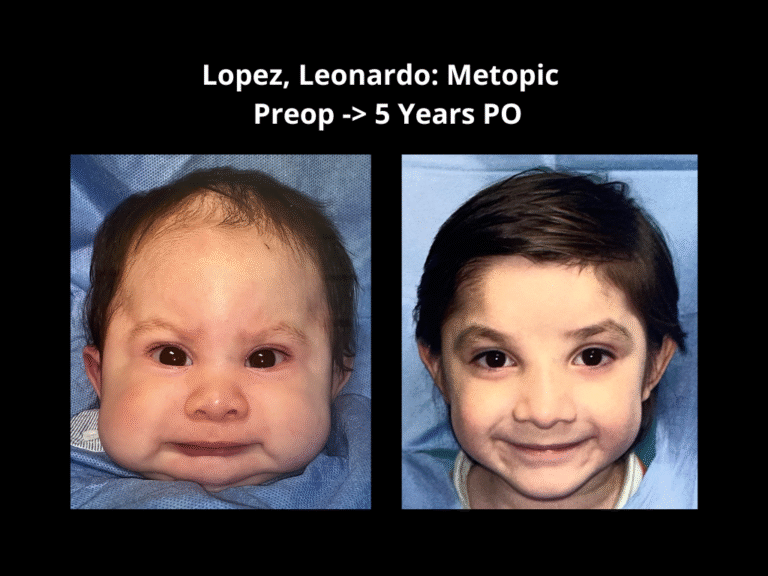
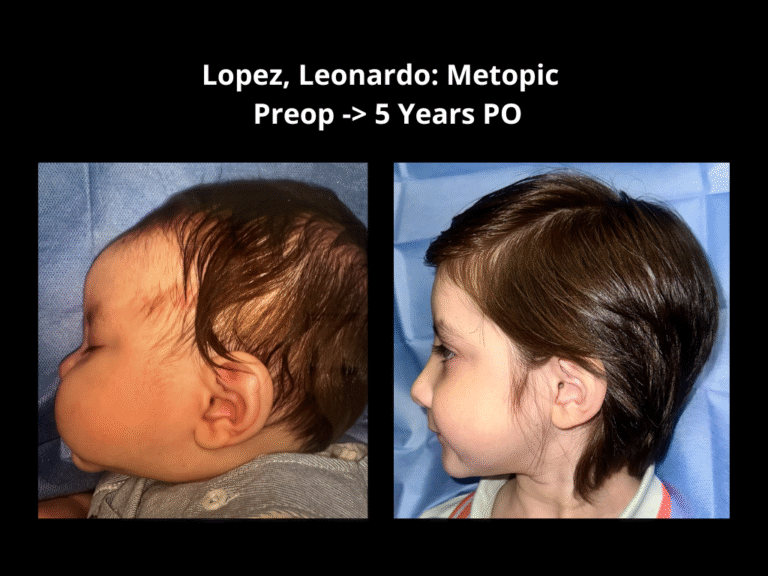
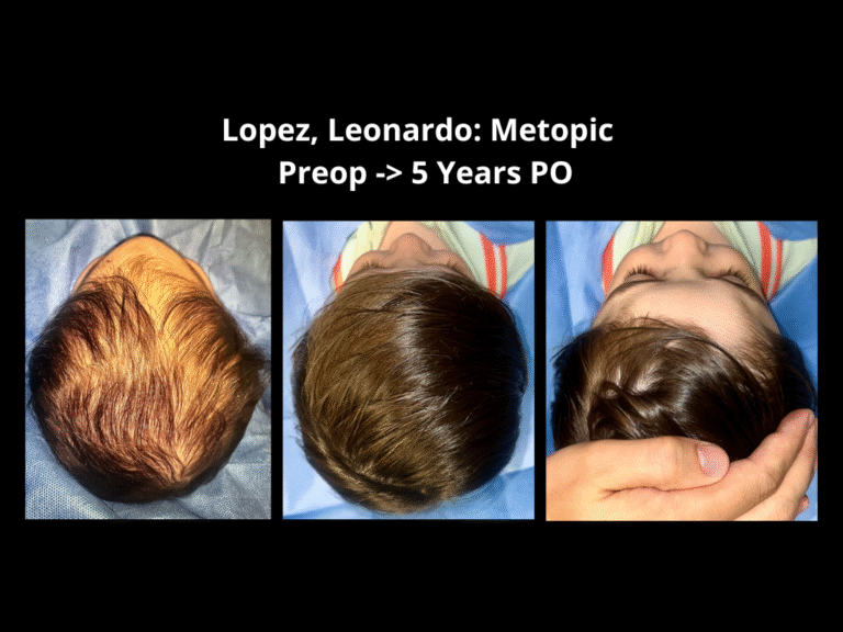
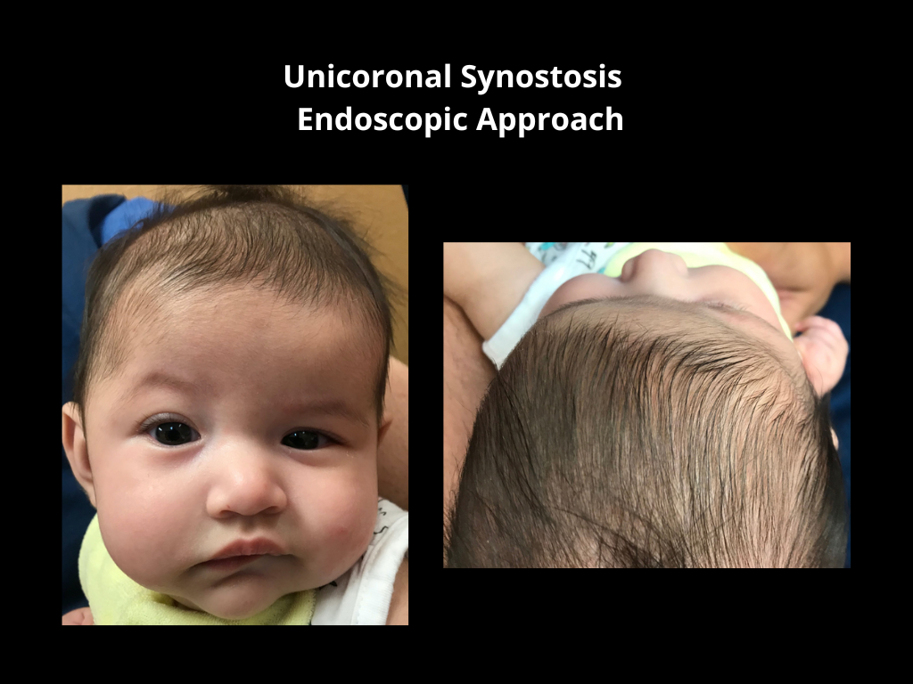
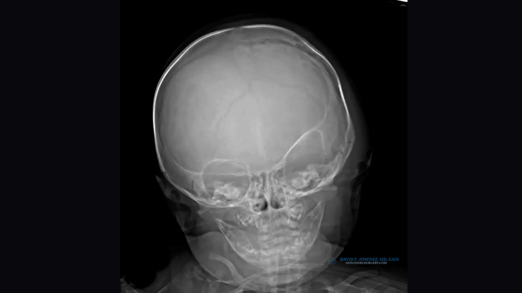
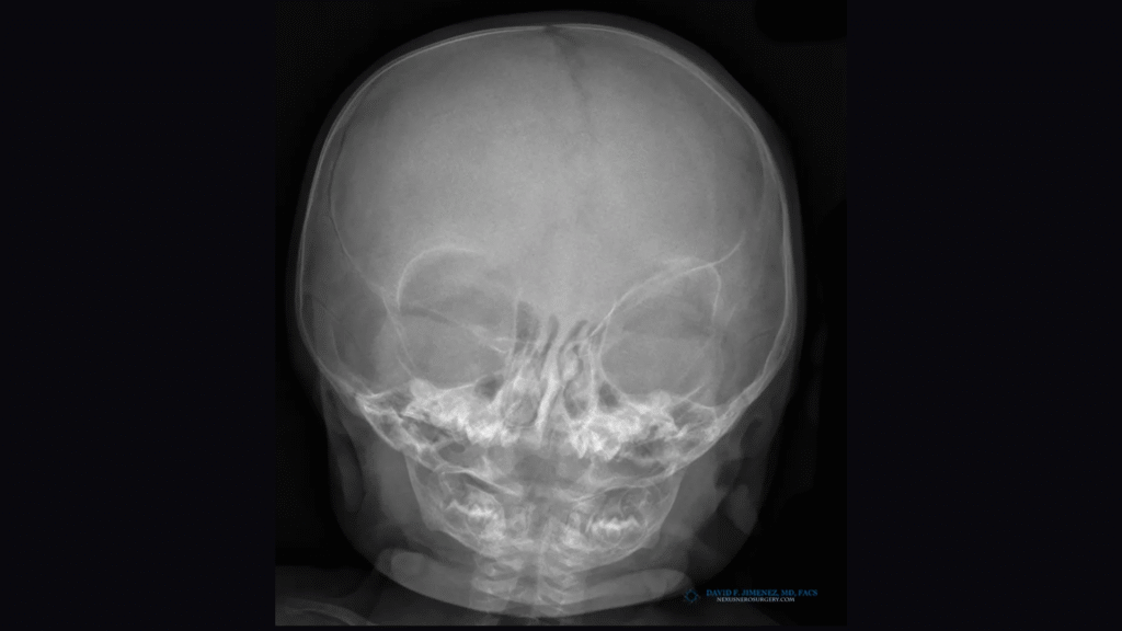
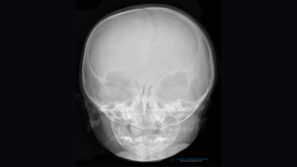
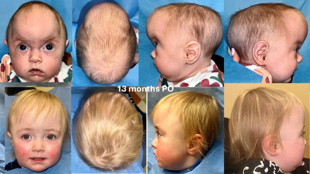
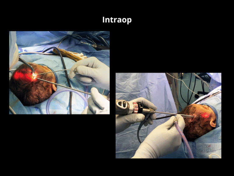
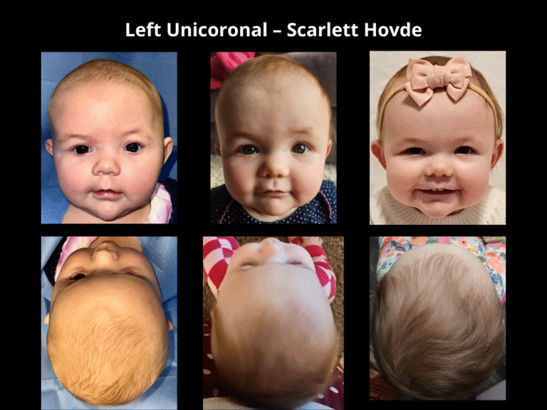
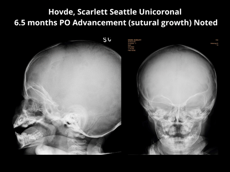
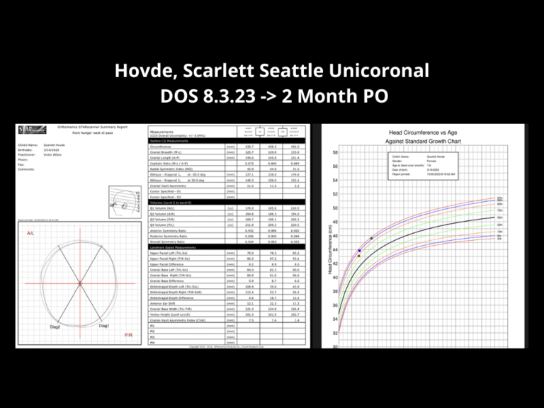
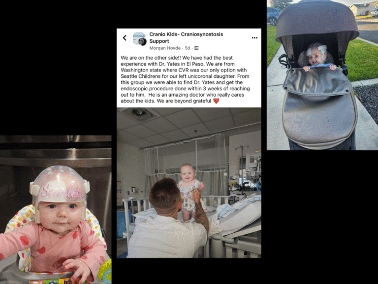
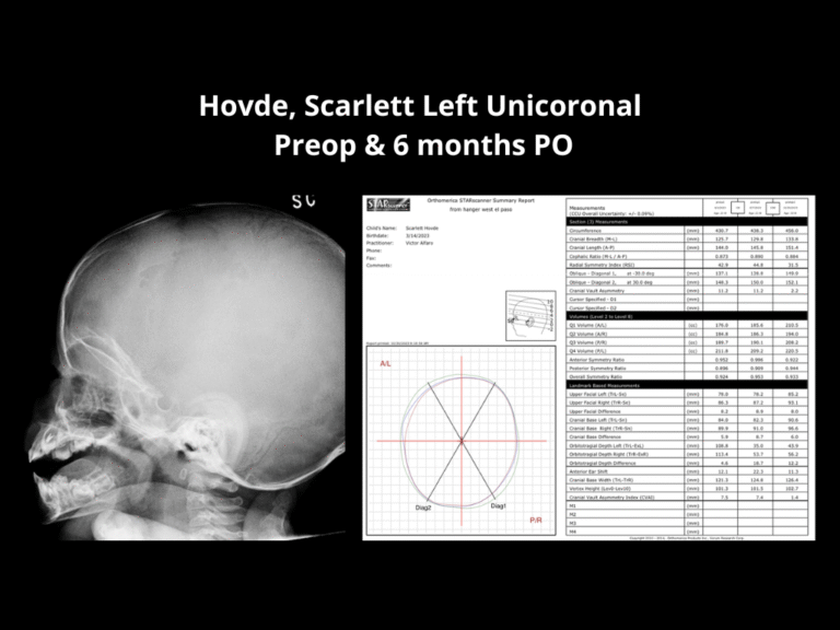
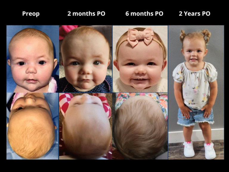
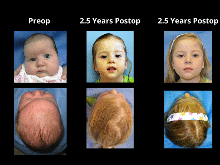
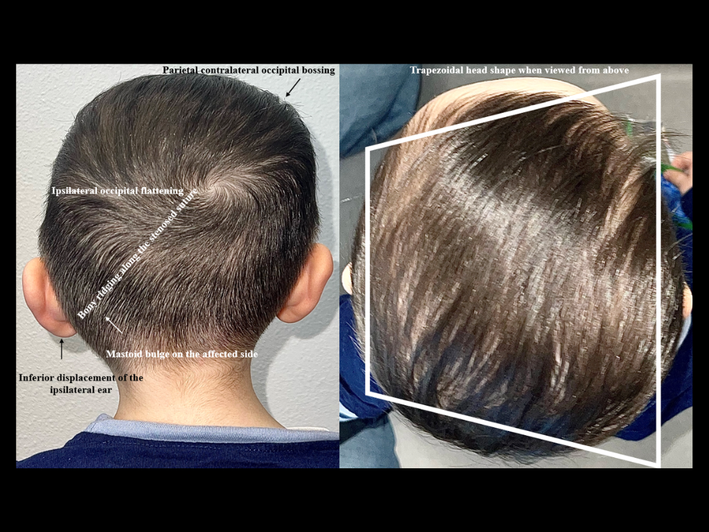
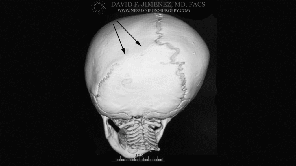
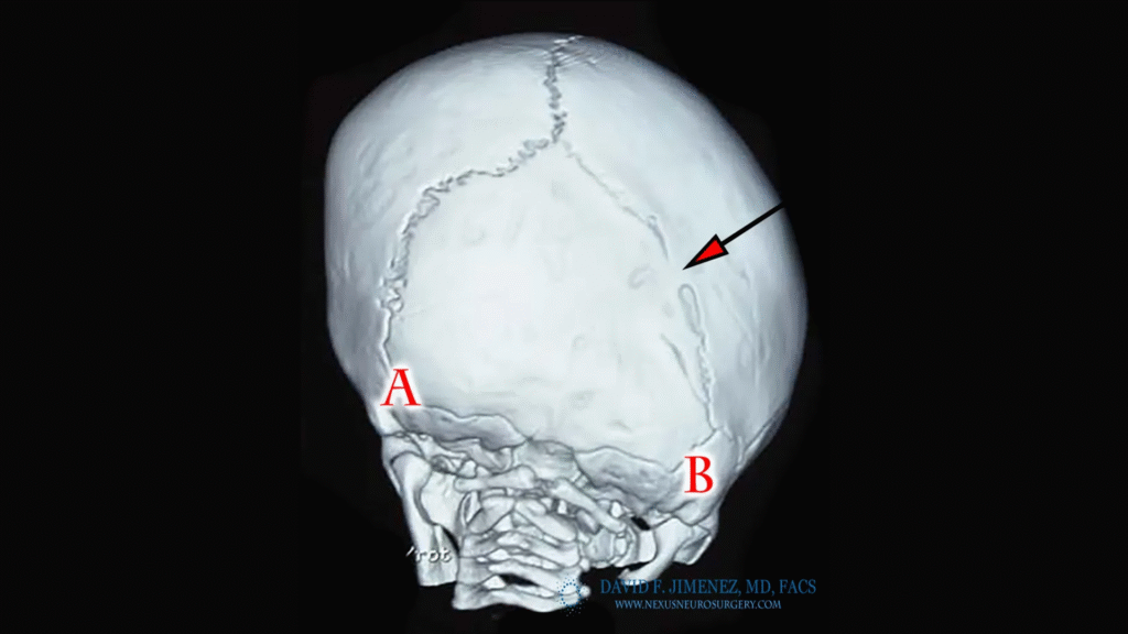
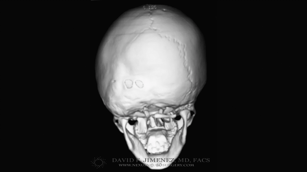
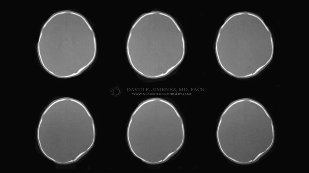
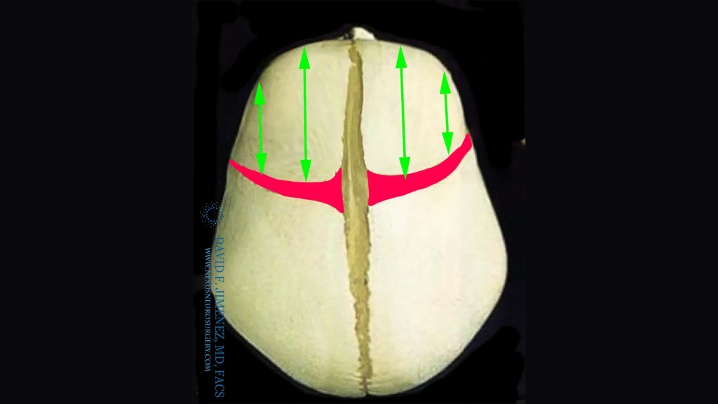
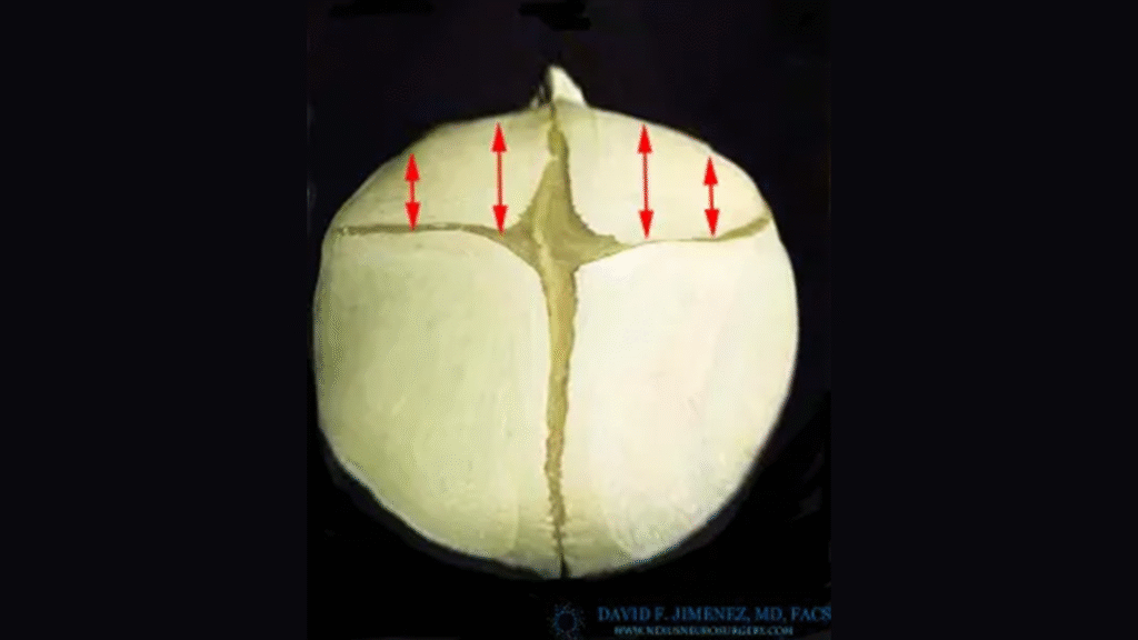
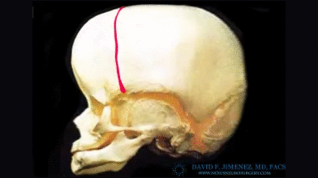
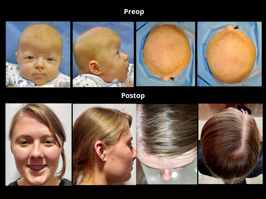
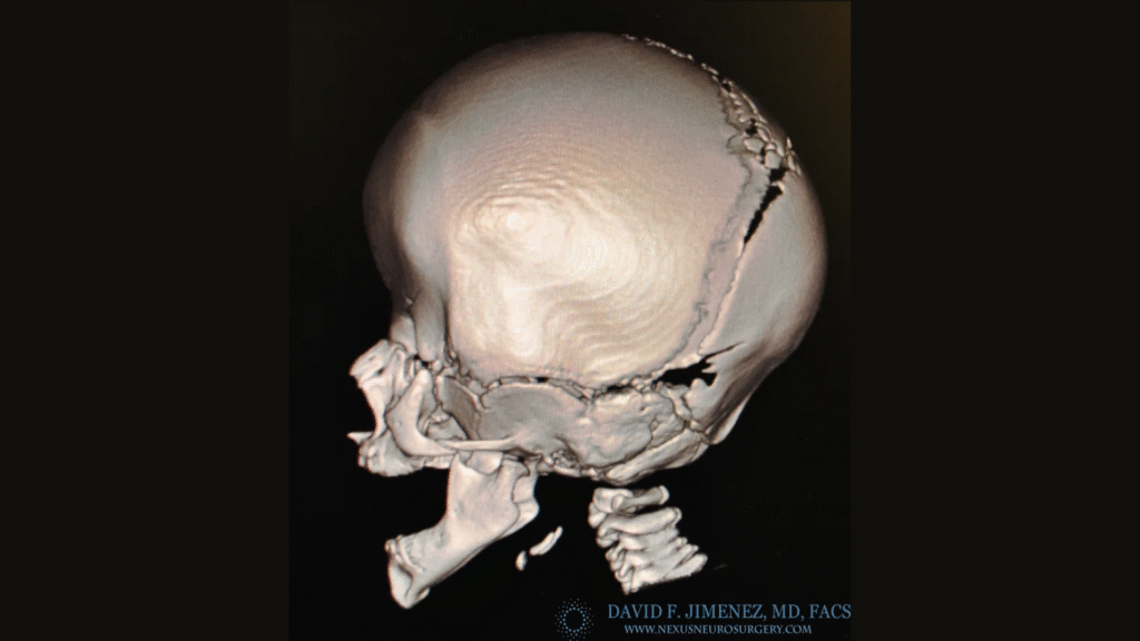
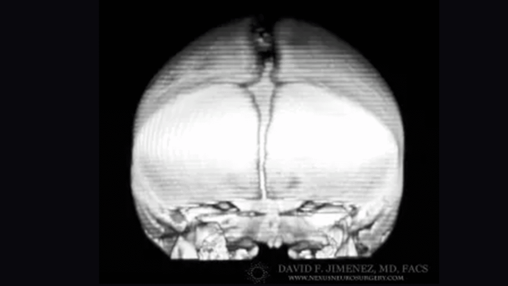
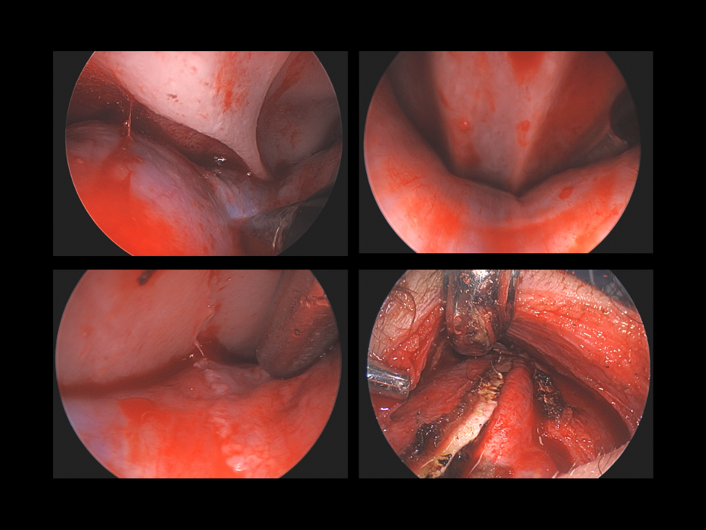
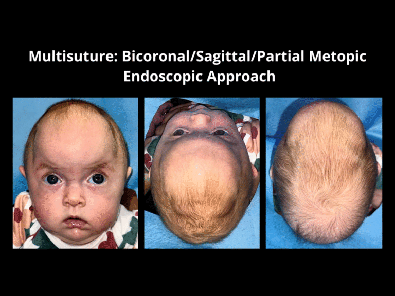
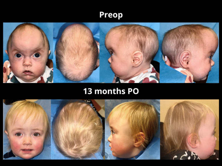
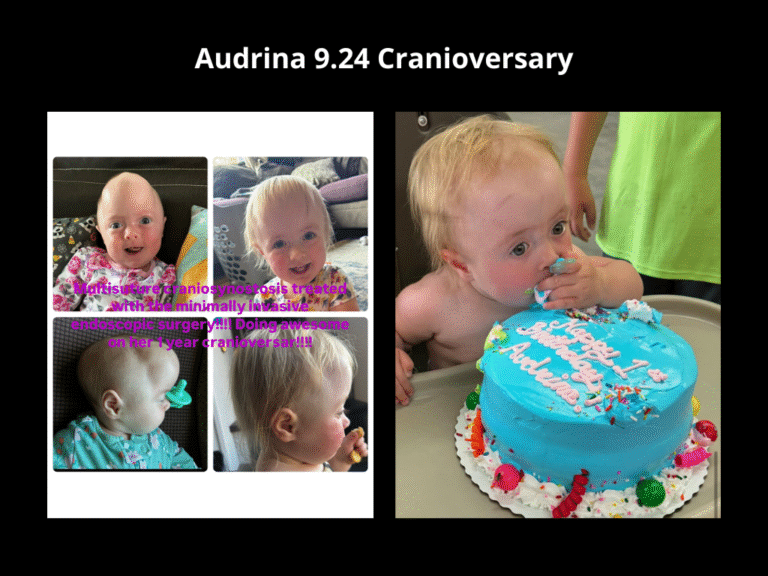
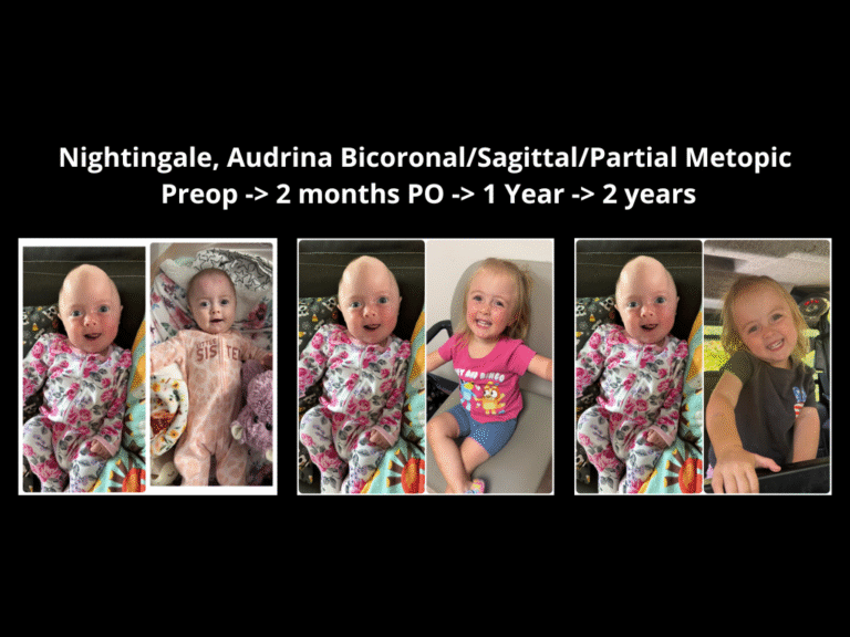
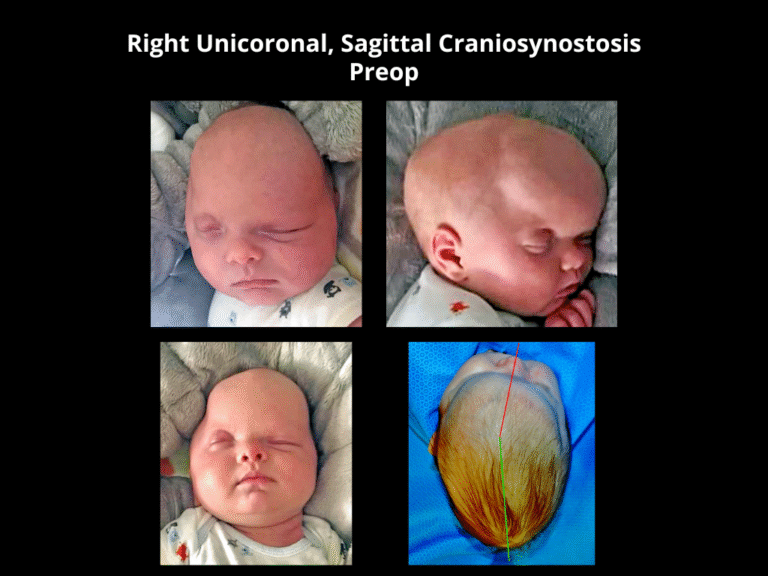
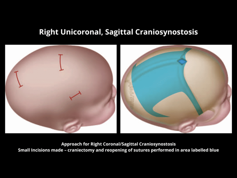
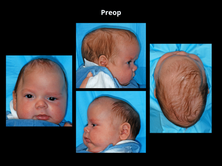
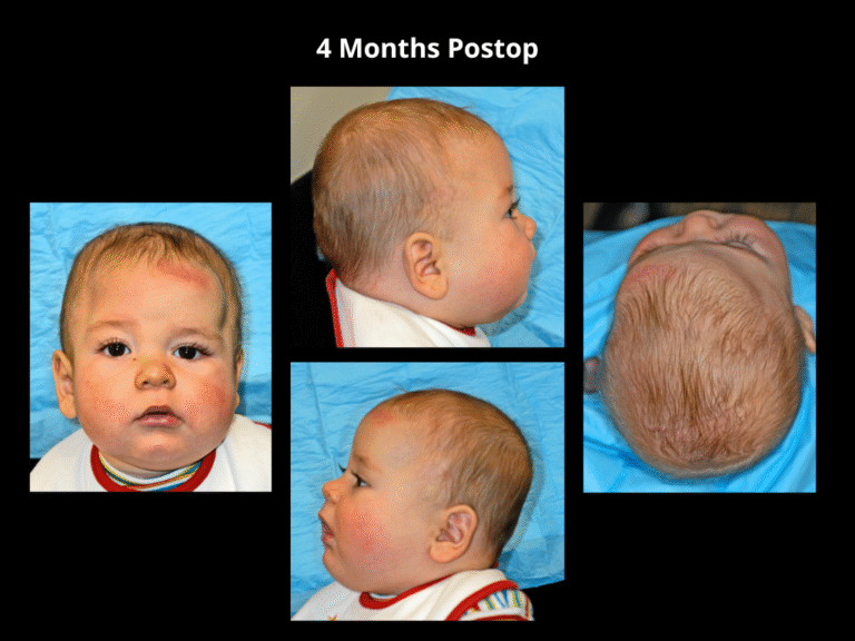
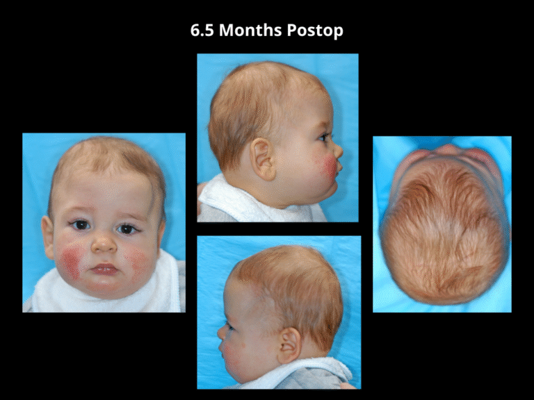
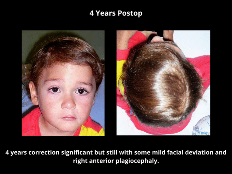
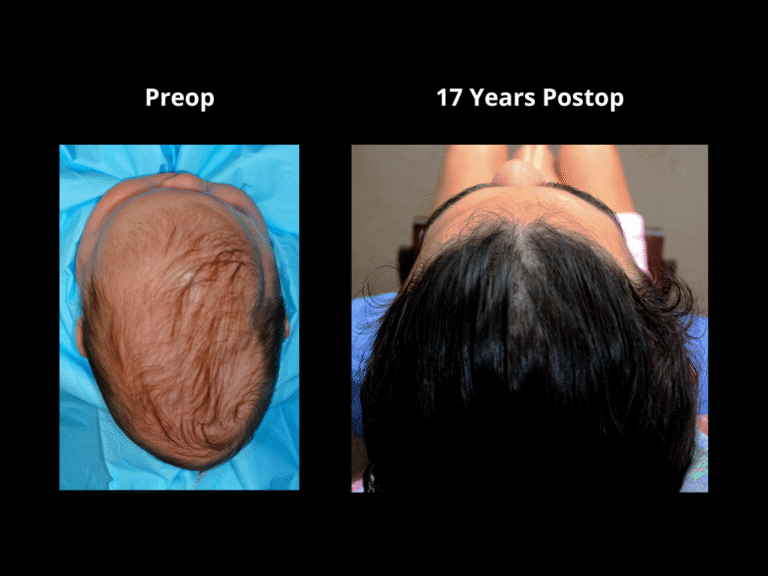
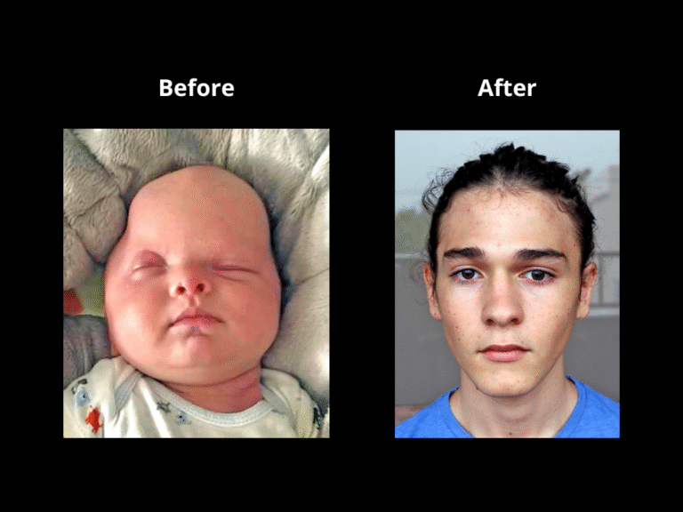
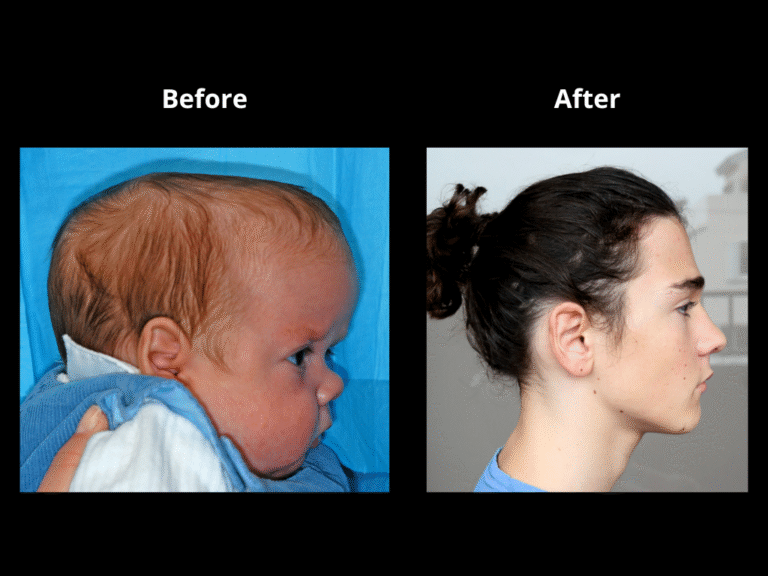
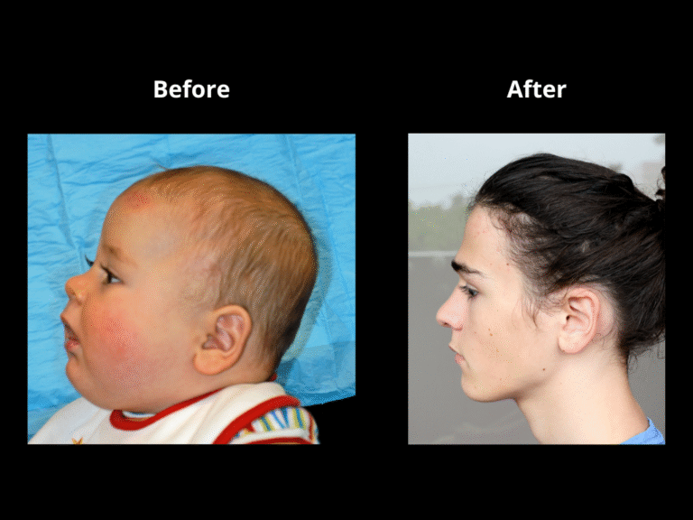
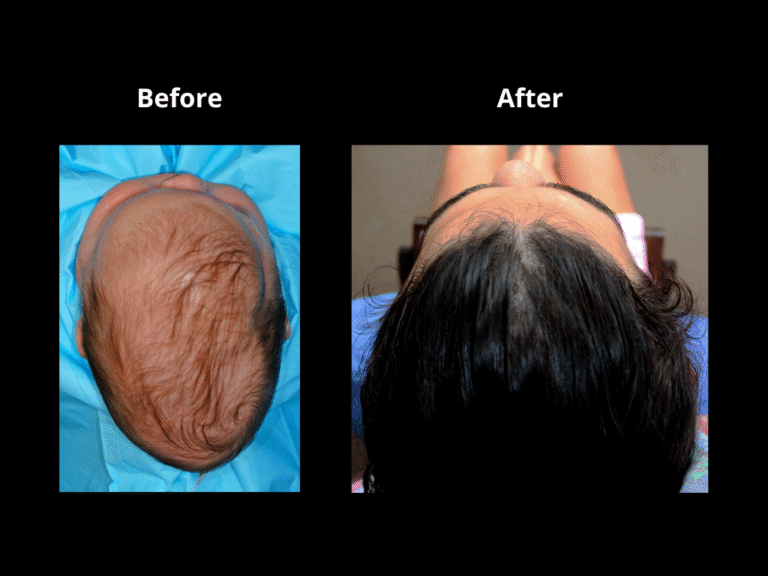
Endoscopic-Assisted Surgery

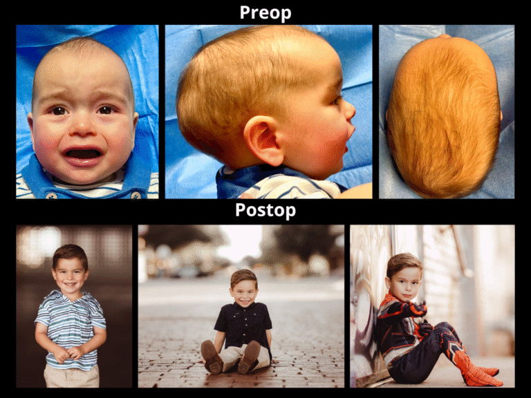
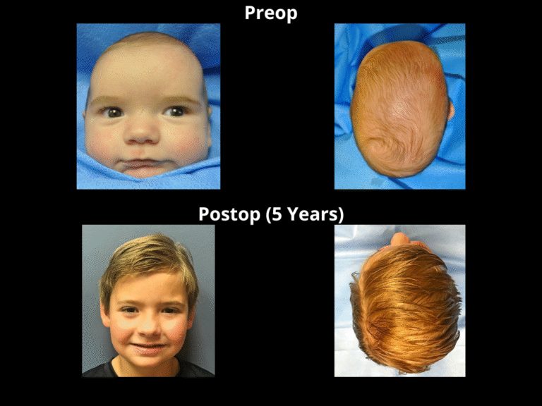
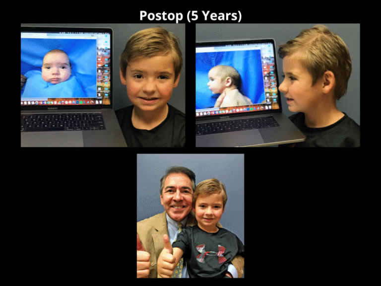
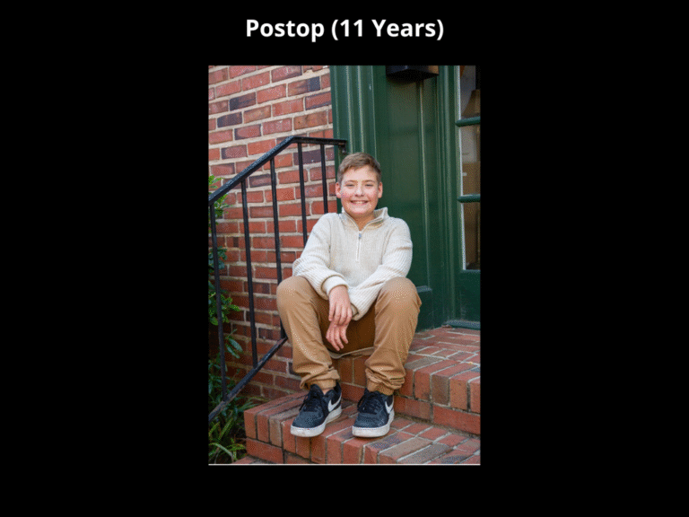
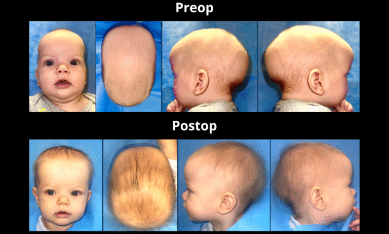
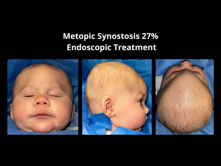
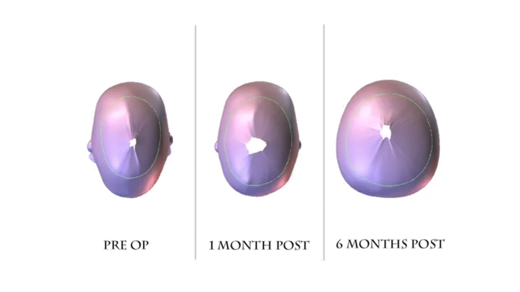
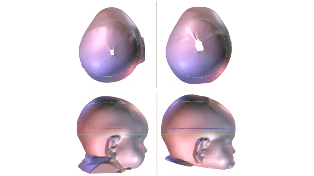
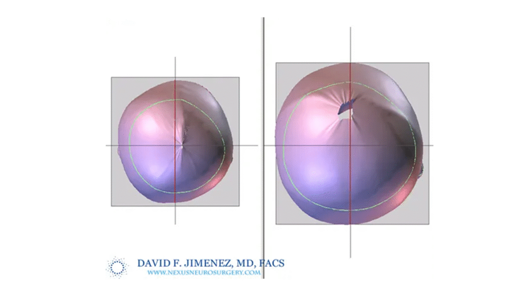
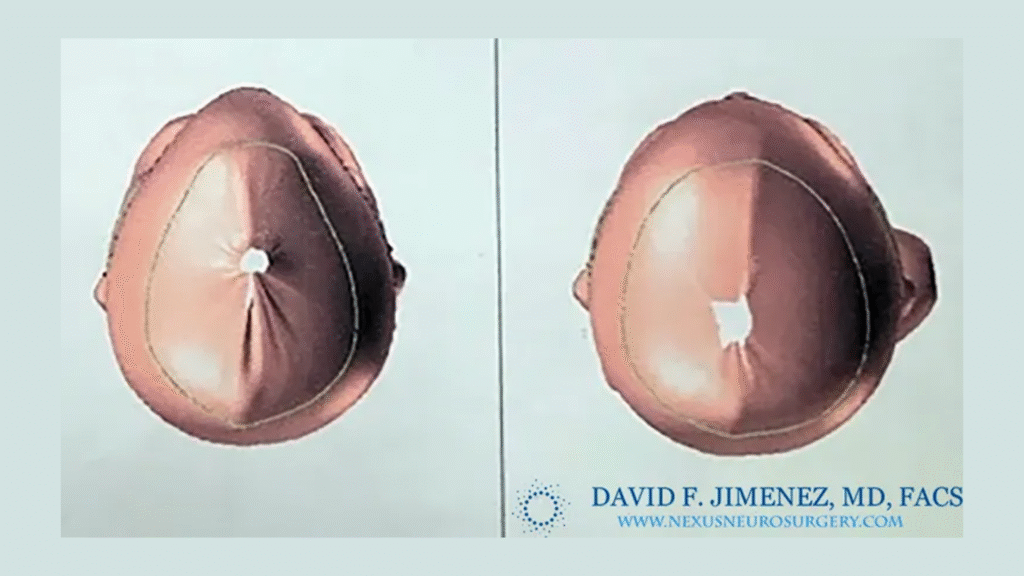
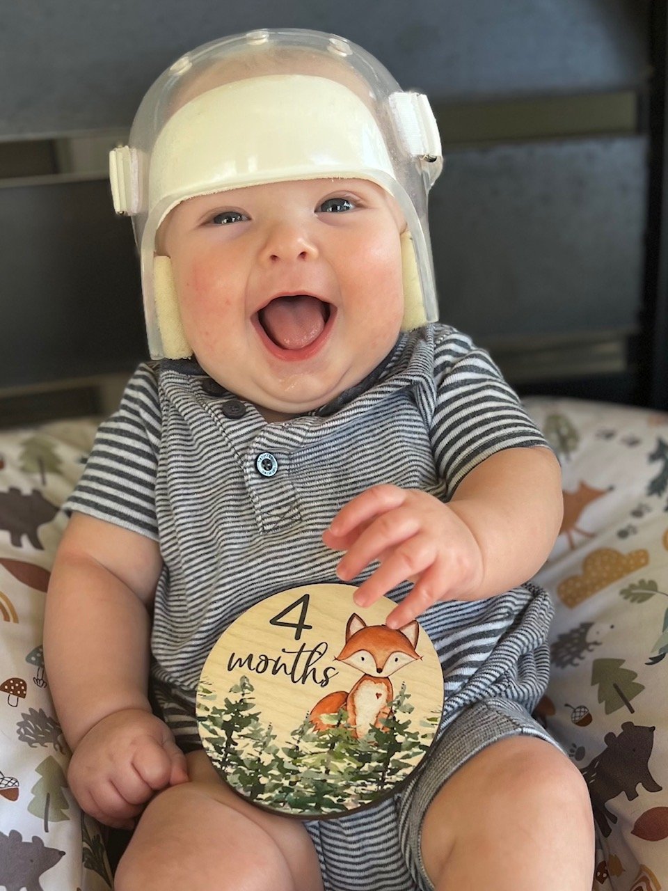
Endoscopic-assisted craniosynostosis surgery is a minimally invasive technique developed to treat infants diagnosed within the first few months of life. Introduced more than two decades ago, this approach allows for safe and early correction of abnormal skull growth by releasing the fused suture through small incisions.

The principle behind the endoscopic method is to use the natural, rapid growth of the infant brain to reshape the skull once the restriction is released. Because the incisions are small and the bone removal limited, this method reduces surgical time, blood loss, swelling, and hospital stay compared to open cranial vault reconstruction.

Procedure Details

Endoscopic-assisted surgery is most effective when performed before 12 weeks of age. The operation involves:

Making two or three small scalp incisions over the fused suture

Using an endoscope to visualize and release the fused suture safely

Removing a narrow strip of bone to allow skull expansion

Relying on natural brain growth to guide skull remodeling postoperatively

The procedure typically takes between one and two hours. Most infants are discharged within 24 to 48 hours after surgery. Recovery is rapid, and infants generally resume normal feeding and comfort within a day.

Sagittal Synostosis

The sagittal suture runs along the midline of the skull, from the soft spot to the back of the head. When it fuses too early, the skull grows long and narrow in a shape called scaphocephaly.

The sagittal suture is located in the midline and behind the soft spot as demonstrated by the red area.

Back view of a normal newborn skull shows the back extension of the sagittal suture as it reaches the lambdoid sutures.

Upon premature closure of the suture, the skull cannot grow in the direction of the red arrows. The normal brain grows in the path of the other open sutures and of least resistance as shown by the green arrows.

The endoscopic approach uses one small incisions along the sagittal suture. The fused bone is carefully removed under direct visualization, creating a narrow strip that allows the skull to widen naturally as the brain grows

Sagittal Synostosis

Before & After

Testimonials

Sagittal Synostosis

Postoperative Course

Most infants experience minimal swelling. Feeding resumes the same day, and comfort returns within 24 hours. The shape improvement becomes evident within weeks.

Metopic Synostosis

The metopic suture extends from the soft spot to the top of the nose. Early closure results in a triangular forehead shape, or trigonocephaly, often accompanied by a midline ridge and close-set eyes.

The metopic suture is located in the midline and in front the soft spot as demonstrated by the red area.

The metopic suture extends from the soft spot all the way down to the root of the nose (nasofrontal suture) in the area between the eyes.

The premature closure of the metopic suture prevents the front center of the skull from moving sideways and the front sides from  moving forwards (red arrows) The midline moves forward ( green arrow) causing a midline ridge and the classical triangular shaped head.

Through one small incision near the forehead, the fused suture is released under endoscopic guidance. The skull is then allowed to expand naturally in the recessed areas.

Testimonials

Before & After

Most infants experience minimal swelling. Feeding resumes the same day, and comfort returns within 24 hours. The shape improvement becomes evident within weeks.

Metopic Synostosis

Post Operative Helmet Therapy

Following surgery, the infant is fitted with a custom cranial molding helmet. The helmet is a vital part of the treatment process. It does not restrict brain growth but gently redirects it, allowing the skull to develop a normal shape.

Helmets are adjusted regularly to accommodate rapid head growth. Therapy generally lasts between six and twelve months. The combination of early surgical release and guided helmet therapy produces predictable, long-lasting results.

Metopic Synostosis

CT Imaging

CT Imaging demonstrating Metopic Synostosis, when obtained, shows postoperative regrowth over the released bone area and complete normalization of forehead contour within two to three years.

Coronal Synostosis

The coronal sutures extend from each ear to the soft spot at the top of the head. When one suture fuses early (unicoronal synostosis), it causes flattening of the forehead, elevation of the eye socket on the affected side, and nasal deviation.

Endoscopic Treatment

A small incision is made above the fused coronal suture. The closure is released endoscopically, permitting balanced forward growth of the affected forehead region.

Coronal Synostosis

The classical presentation consists of coronal craniosynostosis consists of:

1. Elevation of the eye on the affected side (vertical dystopia)

2. Deviation of the nose to the opposite side

3. Flattening of the forehead on the affected side (frontal plagiocephaly)

4. Protruding eye on the affected side (proptosis)

5.  Overgrowth of forehead on the opposite side.

6.  Head deviates to the side (cranial scoliosis)

Coronal Synostosis

Correction of the vertical dystopia and the harlequin sign after endoscopic treatment of left coronal synostosis

Day of Surgery

Two months after surgery

Six months after surgery

Endoscopic Treatment

In selected infants under 12 weeks, both coronal sutures are released endoscopically to restore anteroposterior skull growth and reduce cranial height.

Coronal Synostosis

Before & After

Coronal Synostosis

The coronal  sutures are located on either side of  the head and adjoin the soft spot in the middle. Their function is to allow the frontal lobes of the brain to grow and move forward. This in turn allows the forehead, eyes, eyebrows and nose to also move forward and downward. Premature closure results in a number of deformities related to these structures’ inability to advance normally.

Side view of the newborn skull shows the location of the left coronal suture highlighted in red.

Top view of the skull depicts the  location of the coronal sutures on either side of the soft spot and extending to the sides.

The premature closure of the right coronal suture prevents the front right side of the skull from moving forwards (red arrows) leading to flattening of the right side of the forehead. The brain takes the path of least resistance and begins to grow  more on the left side (green arrows) leading to frontal bossing of the right.

Front view of a newborn skull with right coronal synostosis. The right eye socket is elevated and pulled upwards giving the appearance on skull x-rays of the so called “harlequin eye”.

The nose is pushed to the opposite left side (green arrow), the right eye socket is elevated and moves higher than the left one (red arrow), the right side of the face is pushed inwards (blue arrows) while the left  side of the forehead is pushed outwards (yellow arrows).

Coronal Synostosis

The classical presentation consists of coronal craniosynostosis consists of:

1. Elevation of the eye on the affected side (vertical dystopia)

2. Deviation of the nose to the opposite side

3. Flattening of the forehead on the affected side (frontal plagiocephaly)

4. Protruding eye on the affected side (proptosis)

5.  Overgrowth of forehead on the opposite side.

6.  Head deviates to the side (cranial scoliosis)

Two month old male with significant cranial-facial deviation (scoliosis) as evidenced by the angulation of a line going from the root of the nose to the mid chin and top of the head as seen in coronal synostosis

Cranial scoliosis (red line) and vertical dystopia (left eye elevated above right eye) as demonstrated by the black line in infant girl with left coronal synostosis.

Testimonials

Lambdoid Synostosis

The lambdoid suture runs along the back of the skull. Its early closure results in flattening of one side of the occiput and downward displacement of the ear and mastoid on the affected side.

The lambdoid suture is located in the back of the skull as seen on this newborn’s skull and marked with red.

Both lambdoid sutures, marked in red, as seen on the back of a newborn’s skull.

Back view of an adult skull showing the location of both lambdoid sutures (red lines) as the extend from the mastoid bones to the sagittal suture (black lines) on the top of the skull.

Endoscopic Treatment

A small incision is made over the back of the skull, and the fused suture is released endoscopically to allow symmetrical posterior growth.

Lambdoid Synostosis

Radiology

Lambdoid synostosis can resemble positional molding, so 3-D imaging or CT is often used for accurate diagnosis and postoperative assessment.

3-D CT scan of patient with partial closure of the left lambdoid suture near the midline sagittal suture.(Black arrows)

3-D CT scan of infant with an area of “spot welding” in the middle of the right lambdoid suture. Notice that even this very small amount of  synostosis causes significant deformation of the skull base as seen with downward displacement of the right mastoid bone (B) in relation to the normal mastoid bone (A).

3-D CT scan of patient with premature closure of most of the left lambdoid suture.

CT scan showing thin slices of the skull and a closed left lambdoid suture.

Arrow on the left shows the closed suture whereas the arrow on the right shows an open suture on the other side.

3-D CT scan shows the downward displacement of the mastoid bone in this child with left lambdoid synostosis.

Lambdoid Synostosis

Before & After

Endoscopic Treatment

In infants diagnosed very early, selected multi-suture cases may benefit from staged endoscopic releases to improve cranial expansion while minimizing invasiveness.

Bicoronal Synostosis

Bicoronal synostosis involves premature fusion of both coronal sutures, producing a shortened and widened skull (brachycephaly) with shallow eye sockets and prominent forehead height.

Open coronal sutures (red areas) allow both frontal lobes to grow and advance forward (green arrows).

Closed coronal sutures keep the frontal  lobes from moving forward and cause a shallow and foreshortened frontal cranial vault (red arrows).

Normal brain growth allows the bones around the eyes and face to move forward.

Premature bicoronal synostosis keeps the bones from moving forward and causes the mid face and eye sockets to be recessed and the forehead to bulge abnormally.

Bicoronal Non-Syndromic Synostosis

Radiology

CT imaging confirms the diagnosis and postoperative normalization of cranial shape and proportions.

Multi-Suture Craniosynostosis

Bilateral Lambdoid + Sagittal  Craniosynostosis
“ Mercedes Benz Pattern”

Sagittal & Bilateral Lambdoid

Preop -> 5 Months Postop -> 5 Years Postop

Inside Endoscopic View of

Fused Lambdoid Sutures

Before & After

Before & After

Right Unicoronal Synostosis

It occurs when the growth line (suture) on the right side of a baby’s skull closes too early. This early fusion can cause the forehead and eye area on one side to appear more prominent or uneven. With early diagnosis and proper treatment, most children achieve normal head shape and healthy development.

Right Unicoronal Synostosis

Before & After

Helmets

After surgery, a cranial orthosis (helmet) is used to guide the skull into a more natural shape and address any deformities caused by craniosynostosis. This therapy is generally well tolerated, as reflected in the comfort and smiles of these babies.

The helmet does not restrict or interfere with brain growth in any way. It is carefully designed to guide and support abnormal or compensatory skull growth while allowing the recessed areas to expand into their proper shape. Careful analysis of head circumference growth charts in all of our patients confirms that brain growth occurs normally, with no decline from expected levels. In addition, neuropsychological testing consistently demonstrates that all children develop normally, with no negative effects from helmet therapy.

3-D images show the progressive correction of this infant with sagittal synostosis seen at one and six month follow up.

3-D images of a baby with metopic craniosynostosis and the early improvement seen in a 4 month follow up scan.

STAR scan of a child with left coronal synostosis before (left) and  1 year (right) after endoscopic surgery.

Before and after endoscopic surgery for patient with metopic synostosis.

Helmets

Helmet Photo Gallery

Begin Your Child’s Treatment Journey

Craniosynostosis, whether sagittal, metopic, coronal, bicoronal, lambdoid, or multi-suture, can be treated safely and effectively when identified early.
The El Paso Craniofacial Team offers one of the most experienced endoscopic craniosynostosis programs in the nation, combining advanced minimally invasive techniques with expert helmet therapy to ensure every child achieves a healthy, natural head shape and a bright future.